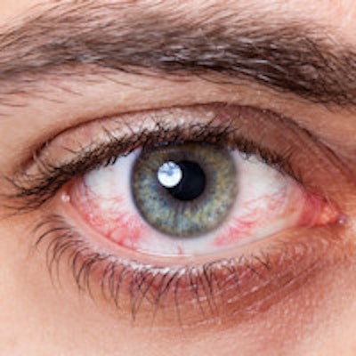
A pioneering exhibit about the role of microscopy coil MRI for the assessment of the orbit and periorbital structures led to a U.K.-Australian team collecting a prestigious Magna Cum Laude award at last month's RSNA 2015 meeting in Chicago.
High-resolution microscopy coil MRI can be added to the diagnostic arsenal with only a little additional, routinely available hardware, and can be used to elucidate deep-tissue involvement and tissue characterization, according to lead author Dr. Nick Dobbs, third-year radiology registrar at Ninewells Hospital in Dundee, U.K. It can assist clinical decision-making about conservative, medical, surgical, or endovascular therapy.
"The complex anatomy of the eyelid makes clinical assessment of deep lesion extent impossible," he noted. "Microscopy-coil MRI allows preoperative planning of the surgical field to minimize the extent of surgery, whilst maximizing clear resection margins. The goal is to keep a functioning eye whenever it is safe to do so."
The emerging technique involves placing a small radiofrequency receiver coil close to the area of interest. The small field-of-view coil results in a high signal-to-noise ratio, producing high-resolution images. It demonstrates the fine detail of small-scale anatomy, and boosts preoperative decisions about lesion involvement of critical structures.
At Dundee, a 1.5-tesla MRI scanner with 32 receiver channels (SQ-engine gradients up to 45 mT/m, slew rate of 200 T/m/s) and a small loop radiofrequency receiver coil (40 mm diameter) are used. T1-weighted and T2-weighted turbo spin echo (TSE) sequences are employed, along with a sub-millimeter pixel size (0.3 mm), slice thickness of 1.5 mm, and no inter-slice gap. Images are acquired in any desired plane, and if required, intravenous gadolinium contrast agent is administered.
| Microscopy-coil MRI sequence parameters | ||
| T1 TSE | T2 TSE | |
| Repetition time | 400 msec | 4,000 msec |
| Echo time | 15 msec | 123 msec |
| Average | 2 | 3 |
| Field-of-view | 80 x 70 | 80 x 70 |
| Matrix | 224 x 256 | 224 x 256 |
| Slice thickness | 1.0 mm | 1.5 mm |
| Turbo factor | 3 | 13 |
| Voxel size | 0.3 x 0.3 x 1.5 mm | 0.3 x 0.3 x 1.5 mm |
| Acquisition time | 5:42 | 7:06 |
To position the coil, the patient must lie comfortably, with his or her head in the posterior portion of the head coil. Headphones can be used to relax and immobilize the patient, and the coil is positioned over the eye. Further immobilization can be achieved with foam wedges. Place a swab under the coil for comfort, and use tape to prevent movement, recommend Dobbs and his colleagues in their e-poster.
In the radiology reports, ophthalmic surgeons want to know about the size, shape, and extent of the structure. They need information about whether the extension is completely extraconal or intraconal, because purely extraconal lesions are more often appropriate for surgery. Also, is the tarsal plate invaded? Resection of the eyelids requires complicated reconstructive surgery, and the surgeon need to know about the relationship to the nasal bone, lacrimal gland, muscles, optic nerve, and globe.
An offer to review the images together can help to optimize patient outcome and build interdisciplinary relationships, the authors noted.
Pitfalls and pearls
"Long acquisition times (7 minutes) may result in movement artifacts. Surprisingly, short involuntary eye movements are averaged out. Persevere!" they advocated. "Lesions extending deeper than the orbital apex may require additional head coil sequences to depict intracranial involvement."
The depth of field is limited in microscopy coil MRI, and when there is pathology deep to the orbital apex, it is important to use both micro and standard head coils. Also, because mascara causes extensive metallic artifacts, make it clear in the pre-examination patient information that makeup must not be worn, Dobbs and colleagues stated.
The technique can successfully be applied in children. Take time to ensure the child is comfortable, and the presence of a family member in the MRI suite can provide reassurance, and an intact dermal fat plane reliably excludes tissue invasion, they added.
Effective use of microscopy coil MRI can clarify deep lesion involvement of the nasal bone and tarsal plate, extending the resection required for clearance, and joint radiological and surgical review of the images helps the surgeon to determine optimal resection margins. While usually most beneficial for depicting structural invasion, the technique also has a role in tissue characterization, the authors continued.
"The high resolution of microscopy coil MRI makes it a great problem-solving tool to elucidate the origin of orbital masses. Better planning maximizes the chance of clear surgical resection margins," they wrote. "Accurate depiction of structural involvement can minimize risk, and sometimes steer a conservative approach. Microscopy coil MRI guides the appropriateness of surgery and alternate endovascular treatment. One of the main advantages is the ability to depict local involvement, to guide surgical management."
Dobbs et al recalled one case of a patient with bleomycin sclerotherapy. The patient said people used to look at him and think he had been in a fight, but following treatment, he and his wife are delighted with the aesthetic outcome.
The technique also has applications in the following areas:
Basal cell carcinoma, which is the most common skin malignancy and is more frequent in older people. It is caused by the stochastic effects of exposure to ultraviolet radiation, which tends to occur in sun-exposed areas. Although benign, they are locally invasive over time (rodent ulcers).
Cavernous malformations, or cavernous hemangioma, which is the most common adult vascular lesion. The patient presents with painless proptosis. The pathology is disputed pathology, being of venous origin or low-flow arteriovenous malformation. They enlarge slowly and progressively, and do not resolve spontaneously.
Capillary hemangioma, which are present from birth and are usually extraconal, but may extend intraconally. They are nonencapsulated lesions with multiple lobules, and most are self limiting. Treatment is with systemic/intralesional steroids, radiotherapy, or debulking. Good reports exist from intralesional bleomycin in refractory cases.
It elegantly demonstrates the anatomy of the levator aponeurosis. The levator palbebrae superioris muscle terminates at the levator aponeurosis, which has two main constituents, contributing to elevation of the eyelid.
The tarsal plate. Each eyelid contains a tarsal plate to add stability, offering a firm base to aid in suspension of the eyelid. Meibomian glands line the tarsal plate, producing an oily material that coats the tear film, reducing evaporation. The oily material offers easy identification of the tarsal plate due to the innate high T1 signal.
It is vital for radiologists to become familiar with microscopy-coil MRI of the eye, appreciate the anatomy of the orbits as demonstrated on the technique, understand the added benefits for surgical planning, review selected cases to facilitate important learning points, and identify pearls and pitfalls, the authors concluded.
The research was conducted in collaboration with radiologists at Gold Coast Radiology in Queensland, Australia, and the University of Wales in Cardiff, U.K., as well as ophthalmologists at Ninewells Hospital.


