Mobile devices can be used as an effective tool to link a radiology department and a forensic pathology institute at two different locations with a common workflow, enabling forensic pathologists to view CT images and reports offline during autopsies, German researchers have found.
 The aim is for the forensic pathologist to have the report and CT data onsite during an autopsy, according to Ignaz Reicht.
In a presentation at ECR 2014 in Vienna, a team led by Ignaz Reicht, researcher at the Software development for Integrated Diagnostic and Therapy (SIDT) unit of the German Cancer Research Center (DKFZ) in Heidelberg, showed how an internally developed mobile application led to the integration of the CT infrastructure of the University of Heidelberg's Institute of Forensic Medicine with the department of radiology at the DKFZ. The result has been well received by both forensic pathologists and radiologists.
"The setup was able to facilitate multicenter radiology in terms of mobile viewing," he noted.
One of the challenges to the project was the limited IT infrastructure in place at the Institute of Forensic Medicine's autopsy site. There weren't any workstations for image viewing, and also no local area network (LAN) or wireless LAN. In response, the DKFZ's department of medical and biological informatics developed an offline-capable mobile system to support viewing of CT data during autopsies at the examination area.
The mobile DICOM image-viewing application, called MITKPocket, allows for offline image processing on tablets. It's available for iOS, Android, and desktop computers.
"[MITKPocket is] also part of the IT infrastructure in our radiology department, but mostly it's used for scientific purposes," Reicht said.
Forensic-radiology workflow
 Forensic pathologist viewing CT data using MITKPocket right before performing autopsy. Interaction with two pairs of gloves (thin nitrile gloves and thicker autopsy gloves). All images courtesy of Ignaz Reicht.
The institutions' new forensic-radiological workflow model begins with a CT scanner acquiring forensic images at the Institute of Forensic Medicine. Next, the images are transferred via a virtual private network (VPN) connection to the MITKPocket server located in the radiology department at DKFZ. A forensic pathologist can then download the images from the server to a tablet device.
"On the next day, he can then go into the autopsy and view the images during the autopsy," he said.
In addition to being available via the temporary cache data storage provided on the MITKPocket server, the forensic images are also sent to a dedicated scientific PACS (Chili). Radiologists can read the images either using the MITKPocket application or with the Chili PACS workstation software. The radiologist's report is then sent to the forensic pathologist for access via MITKPocket.
"We reached the aim of this project that the forensic pathologist had the report and the CT data onsite during his autopsy," Reicht said.
Benefits of the setup
The system's technical implementation was very cost-effective, as no major investments were needed, he explained. The researchers also performed a study to determine if MITKPocket was meeting the requirements of forensic-radiological workflow.
After implementation, "MITKPocket was carrying most of the workflow and it served as an appropriate solution," Reicht said. "It showed actually 100% availability in a three-month timeframe."
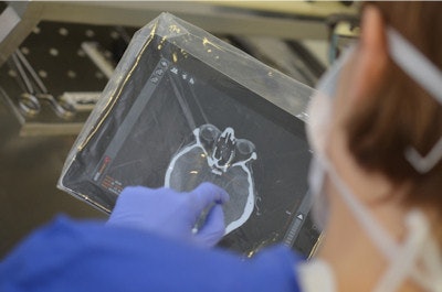 Forensic pathologist viewing CT data at autopsy site using MITKPocket. Interaction with two pairs of gloves (thin nitrile gloves and thicker autopsy gloves) and a protective covering for iPad (transparent singular use plastic bag).
The forensic pathologists realized a number of benefits from the system, he continued. "Since forensic science is low on radiological equipment, this workflow actually provided them access to the radiological infrastructure," he said. "In that case, they can view images offline at the autopsy site."
The radiology department also found the system offered several advantages, including the ability to conduct detailed analysis of workflow and to experiment with modifications, Reicht said.
"From that knowledge, we can put it into clinical workflows," he said.
In addition, radiologists have gained increased access to postmortem images.
"This workflow shows that forensic pathology is a valued partner for research in radiology," he concluded.


