In this Mobile App Spotlight, we're taking a look at CT Anatomy, an iOS app that offers a cross-sectional guide of normal anatomy as seen on CT. It includes more than 350 axial CT views with anatomy, as well as more than 700 anatomical structures along with descriptions, according to developer iCat Medical Software, a U.K.-based mobile app firm.
We chatted recently with George Michalopoulos, a software developer and iCat's CEO, to learn more about CT Anatomy.
AuntMinnieEurope: Could you share some background on iCat Medical Software, the company that developed CT Anatomy?
 George Michalopoulos, CEO of iCat Medical Software.
Michalopoulos: I started developing apps in 2009 as a side project and released my first app, iRad CT, in 2010. Soon, I realized that this side project was taking more and more of my time to maintain and move forward. So in 2011, iCat Medical Software was born. Four years forward, our team now consists of radiologists, radiologic technologists, software engineers, and graphic designers with many years of experience in their fields.
What inspired you to create CT Anatomy? What problem does the app solve or help address?
At iCat, we develop apps we love to use ourselves. And this is what has driven us from the beginning when we developed our first app. We want our apps to have a real-world practicality and application. We thought that displaying drawings or illustrations of the human anatomy has no practical use in the day-to-day work environment -- you're never going to see any illustrations in real-life CT scans!
CT Anatomy helps educate the user on normal human anatomy as seen on the CT. By learning normal CT anatomy, you are taking the first step toward correct diagnosis.
Where do you get the content for the app?
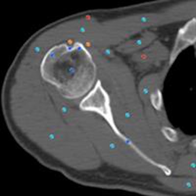
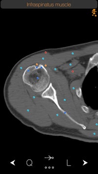 Normal infraspinatus muscle as shown on the CT Anatomy iOS app. Users can tap the dots for more information about each specific anatomical area. All images courtesy of iCat.
The CT images used in the app come from nonpathological CT scans. At first we thought of using images from the [U.S. National Library of Medicine] Visible Human Project, and we acquired the relevant permission to do so. However, we soon realized that the Visible Human Project did not contain all parts of the human anatomy, and we wanted our images to have a consistency and continuity. So we decided to go with the option of using real patient scans. Obviously, we were very careful in obtaining copyright and ethical clearance from the patients and the hospital we worked with.
Who is the target audience for CT Anatomy, and how do you envision it will be used?
Our primary target, of course, is medical professionals -- especially radiologists and radiologic technologists -- but anyone with an interest in cross-sectional anatomy can use and benefit from the CT Anatomy app. The volume of information in regards to the anatomical structures described and the ease of use in the app goes beyond the strict use of it in just the medical sector.
Many universities are already using CT Anatomy for training purposes by using the QuizMe mode in the app. I'm sure radiologists and other medical professionals will use the app in ways we didn't imagine when we started developing it. For example, we had an email from a radiologist who is using the app for educating patients and showing them what normal anatomy looks like.
What are the app's most important features?
A CT Anatomy app can't deviate much from its intended purpose, which is to demonstrate the anatomy of the human body. It has to be complete and comprehensive. However, sometimes a single anatomical name does not give you the whole picture of that structure. So we have taken this one step further by including information in a very easy and nonobstructive way about the anatomical structure you are searching for.
Another feature that I personally like a lot is what we call FollowMe mode. Looking at a 2D image and trying to figure out where an anatomical structure comes from or goes to is sometimes difficult. With CT Anatomy, the user can follow an anatomical structure through its course and understand better its relationship with other adjacent structures.
 Users can access detailed descriptions of anatomy, as seen here in the description of the infraspinatus muscle.
How many users do you currently have for the app?
Since we launched CT Anatomy in 2011, we have more than 10,000 combined downloads (the CT Anatomy app can be downloaded individually or as part of a bundle) in more than 120 countries around the world. We are extremely pleased with the reception of the app by our users, and the feedback so far has been very positive, with five-star reviews on the App Store.
What are your future plans for the app? Are there any new features or content in the works? Do you have any plans to develop a version for Android?
We are always focusing to provide the best for our users. We always listen to what they say and we are preparing a major update with more anatomical structures and more detailed images in 2016. We are also considering adding coronal and sagittal views in future releases of the app.
Our app is already localized in Spanish, and we are looking into expanding to other languages too. Android is not in our immediate future plans.
What other radiology-oriented apps have been developed by iCat?
At iCat Medical Software, we have a product range of 10 apps. Each one of them comes with its own dedicated management, development, and consulting team to ensure accuracy of the information provided. We have developed a series of educational and referencing apps for radiologic technologists (iRad Xrays, iRad CT, and iRad MRI), and along with the Xray Anatomy app, we have a pocket guide for radiologists (iRadiologist - Brain) with the most common brain conditions, with differential diagnoses, analytic details, and images of every pathology described.
We also have some pet projects -- namely, an innovative cranial nerve map (nTube), an MRI simulator (MRI Sim), an estimated glomerular filtration rate calculator (iRad eGFR), and a semiautomatic cardiothoracic ratio tool.
CT Anatomy can be downloaded for $9.99 from the iTunes App Store.


