Low-dose digital tomosynthesis (DT) of the thorax administers a similar radiation dose to plain radiography but is far more accurate, can be applied to most lung nodule evaluations, and is useful for follow-up of the size change of nodules or other lesions, according to new prize-winning research.
On the flip side, low-dose DT produces more noise and has only limited penetration in thick or dense regions. Moreover, it has limited contrast sensitivity to depict ground-glass opacity (GGO) or subtle consolidations, and the scan time of 7-12 seconds is relatively long compared with plain radiography, making the technique vulnerable to motion artifacts, noted Dr. Hong Sik Byun and colleagues from the department of radiology, Samsung Medical Center, Sungkyunkwan University School of Medicine, Seoul.
"If the patient breathes during the scan, back projection will fail and ghost artifact will appear," they stated in an e-poster that received a certificate of merit during RSNA 2012 in Chicago. "For some minor movements, we cannot easily perceive the motion artifact. In these cases, indicators of movement are double rib contours and/or rippling of diaphragmatic dome."
DT is a form of limited-angle CT that allows reconstruction of multiple section images from a set of projection data acquired over a limited range of x-ray tube angles. It offers the potential for improved diagnostic performance over conventional radiography by substantially reducing the visual clutter of the overlying anatomy. Although it does not have the depth resolution of CT, DT provides high-resolution images in the coronal or sagittal plane with a substantial reduction in the radiation dose compared with CT, the authors explained.
Comparison of digital tomosysnthesis (DT) to digital radiography (DR) and CT
Source: Digital tomosynthesis of thorax, Dr. Hong Sik Byun, e-poster presented at RSNA 2012. |
As the chart shows, DT is positioned between DR and CT. The authors note the DT equipment is based on a digital radiography unit, the cost of a DT examination is similar to that of DR (at least in Korea), the radiation dose is also much less than CT, and the reading time and PACS storage size are more similar to CT than radiography.
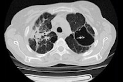
Low-dose DT can show honeycomb fibrosis or dense pneumonic consolidation in patients with usual interstitial pneumonia (UIP). An air bronchogram is well noted in the consolidation of the right upper lobe. However, low-dose DT cannot demarcate the area of GGO in the basal lung because of poor contrast sensitivity, and it is hard to distinguish small nodular consolidation from thick fibrosis on low-dose DT, the researchers pointed out.
For lung cancer screening, chest x-rays (CXR) are useless, while DT is accurate for nodule detection but of limited use for GGO lesions and part solid nodules. CT is more appropriate, and because patients tend to be older and malignant disease is common, the higher exposure is justified.
For tuberculosis screening, CXR are used but are of limited value, and DT is more useful than CXR. CT is accurate but inappropriate because of the relatively young age of patient, the fairly high incidence of tuberculosis in women, and the incidence of benign disease. For benign or chronic disease, radiation exposure comparable to plain radiography is justified, they commented.
The same group has also investigated the use of low-dose DT for the evaluation of the paranasal sinus (PNS) and compared its diagnostic accuracy with a PNS radiography series. For more details, click here.

