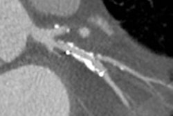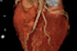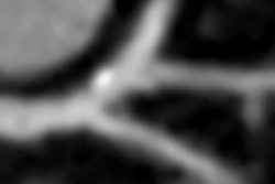
MUNICH - False-positive diagnoses in coronary CT angiography (CCTA) are most frequently caused by regional vessel calcification and motion artifacts, German researchers noted on Monday at the European Society of Cardiology (ESC) annual meeting.
Furthermore, the significantly higher image noise in misclassified CT datasets emphasizes the important role of image quality for a correct identification of significant stenoses in CCTA, according to lead presenter Dr. Jasmin Eisentopf, from the department of cardiology at the University of Erlangen.
"Coronary CT angiography has demonstrated high diagnostic accuracy for noninvasive detection of coronary artery stenoses. However, false-positive diagnoses occur, and the underlying reasons for misclassification have not yet been systematically evaluated," she noted.
For the period between January 2007 and October 2011, the Erlangen team retrospectively analyzed datasets of 571 consecutive symptomatic patients (414 men, 157 women) with an intermediate likelihood for coronary artery disease. These patients showed at least one significant stenosis (> 50% lumen reduction) in CCTA and were referred for invasive catheterization.
The researchers assessed the number of false-positive diagnoses in CT angiography versus invasive angiography, as well as the respective coronary artery. The subjective underlying reasons for inaccurate interpretation were evaluated using four classifications: calcification, motion artifact, image noise, and small vessel diameter. They compared datasets with correct and incorrect interpretation with respect to differences in mean heart rate, body mass index (BMI), Agatston score, image noise, and effective radiation dose.
Of the 571 datasets, 48 (8%) patients (36 male, 12 female) and 128 (14%) coronary arteries showed at least one false-positive diagnosis in CCTA. Of these 128 coronary arteries, two were left main, 41 were left anterior descending, 48 were left circumflex artery, and 37 were right coronary artery. False positives were caused by calcification in 72 cases, motion artifact in 38 cases, and image noise in 10 cases, and were attributable to a small vessel diameter in seven cases.
On a vessel-based analysis, mean heart rate, BMI, Agatston score, image noise, and effective radiation dose for datasets with accurate interpretation and datasets with inaccurate interpretation, respectively, were 61 ± 10 and 61 ± 12 beats per minute, 27.7 ± 4.3 and 28.3 ± 4.9 kg/m², 497 ± 827 and 607 ± 592 (p = 0.030), 27 ± 8 and 31 ± 12 Hounsfield units (p = 0.016), and 8.7 ± 5.1 and 9.6 ± 7.5 mSv.
