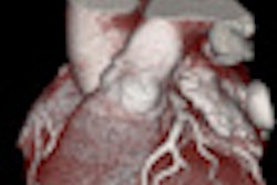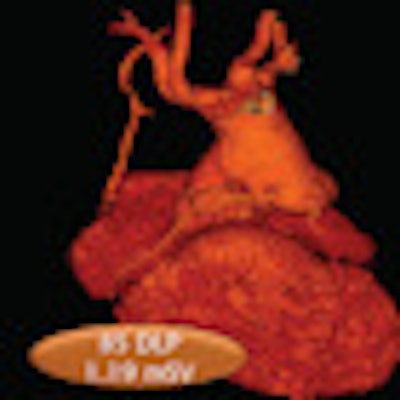
Austrian radiologists have found a way to cut the radiation dose of coronary CT angiography (CCTA) studies to as low as 1 mSv by using a high-pitch scanning protocol on a 128-slice scanner for patients with low heart rates.
Traditional retrospectively gated 64-slice coronary CTA is associated with relatively high radiation doses of approximately 12 mSv, but an emerging high-pitch CT angiography technique, performed on a 128-detector-row dual-source scanner, can reduce dose significantly, said Tobias DeZordo, MD, from the Medical University of Innsbruck. DeZordo presented his group's research on the subject at the 2010 RSNA meeting in Chicago.
The study aimed to assess the performance of the high-pitch technique by comparing it to two other CT protocols -- adaptive sequential prospective electrocardiogram (ECG) triggering and retrospective ECG-gated low-pitch spiral CTA -- in different groups of patients who were triaged primarily by heart rate.
DeZordo, along with lead researcher Gudrun Feuchtner, MD, and colleagues, acquired all images on a 128-detector-row dual-source CT scanner (Somatom Flash, Siemens Healthcare, Erlangen, Germany). The high-pitch Flash protocol uses a pitch setting of 3.4, prospective ECG synchronization, and a short scan time of only 0.3 seconds, he said.
The study population consisted of 103 patients referred to cardiac CT, including:
- 67 patients for coronary CTA
- 7 patients for acute chest pain/emergency department triple rule-out
- 9 patients for coronary artery bypass graft (CABG) surgery or follow-up
- 9 patients for preinterventional planning (transcatheter aortic valve transplantation, totally endoscopic coronary artery bypass surgery, chronic total occlusion)
- 8 patients for pulmonary vein ablation
- 2 patients for evaluation of congenital heart disease/valve assessment
Patients were triaged into one of three protocols based on heart rate:
- High-pitch Flash sequence for patients with heart rates < 60 bpm
- Adaptive sequence scanning for patients at 60-80 bpm and regular sinus rhythm
- Retrospective ECG gating for patients with > 80 bpm or arrhythmia
In addition to variations in the scan protocols, when images are acquired using the adaptive protocol, the scanner software automatically widens the pulsing window based on heart rate, DeZordo said:
- < 65 bpm: 70%
- 65-70 bpm: 40%-70%
- > 70-80 bpm: 30%-70%
Image quality of coronary arteries in coronary CTA was scored as "diagnostic" or "nondiagnostic" (per the American Heart Association's 16-segment classification). Radiation dose was estimated based on dose-length product, while contrast agent volume in CCTA was compared between the three techniques. Among the 103 patients, Flash high-pitch scans were acquired in 45% (n = 46), adaptive sequence in 37% (n = 39), and retrospective ECG gating in 8% (n = 19).
Among the total 67 coronary CTA studies, the triaged protocols included high-pitch Flash scans in 25% (n = 17), adaptive triggering in 52% (n = 35), and retrospective spiral scans in 22% (n = 15). The total mean dose for all protocols for the 67 studies was 4.8 mSv ± 4.7, DeZordo said, but mean doses for the protocols as well as the contrast dose varied, with significantly reduced dose and contrast utilization in the Flash scans.
Radiation dose and contrast by CCTA protocol
|
||||||||||||||||
| p < 0.001 |
In four patients (0.5%), high-pitch CTA had nondiagnostic image quality for at least one coronary artery (right circumflex artery, n = 4; circumflex, n = 1). In each of these patients, heart rates increased during the CT scan to higher than the 60 bpm threshold for optimal Flash scanning (captured heart beats: 60, 61, 61, and 63 bpm). When this occurred, a sequential (n = 3) or spiral (n = 1) exam was appended to ensure diagnostic image quality, DeZordo said. The total radiation exposure in these patients was a mean 3.9 mSv (representing the additional sequence) and 13.5 mSv (representing the additional spiral acquisition).
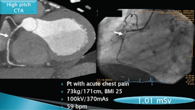 |
| Coronary artery stenosis is detected by coronary CTA (left) and conventional angiography (right) in a patient with acute chest pain. All images courtesy of Gudrun Feuchtner, MD. |
"High-pitch Flash mode scanning is reliable for coronary CTA if the heart rate is lower than 60 bpm, and high-pitch scanning allows for low radiation dose as well as [lower] contrast agent volume," DeZordo said. "High-pitch spiral mode can be integrated in clinical cardiac CT practice using our proposed triage, and permits a tenfold radiation dose savings and contrast volume savings."
Responding to a question from the audience, DeZordo said triage by heart rate only was generally successful; however, very large patients would not be scanned using the high-pitch Flash protocol. The job of the technologist can be complicated, he added, owing to the use of so many different protocols.
By Eric Barnes
AuntMinnie.com staff writer
January 17, 2011
Related Reading
Less DNA damage seen after high-pitch coronary CTA, April 9, 2010
High pitch equals low dose in coronary CT angiography, March 4, 2010
High-pitch DSCT offers speedy images equivalent to slow scans, November 30, 2009
High-pitch cardiothoracic CTA skips breath-holds and high doses, July 7, 2009
Dual-source CT edges into cardiac SPECT turf, March 6, 2008
Copyright © 2011 AuntMinnie.com
