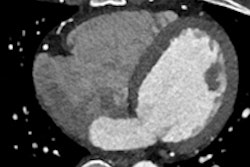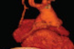
The combination of 256-detector-row CT and iterative reconstruction can reduce dose significantly even in hard-to-evaluate coronary artery bypass grafts (CABG), according to an Italian study.
Patients evaluated using a low-dose ECG-gated coronary CT angiography (CCTA) protocol and iterative reconstruction were scanned at less than half the radiation dose of those given a standard protocol, according to the researchers from Italy. At the same time, image quality didn't suffer, nor did diagnostic accuracy, with the reduced-dose protocol.
"CTA of CABG in comparison with evaluation of the coronary arteries requires acquisition of wider scan coverage, leading to an increase in scan time and radiation dose," noted Dr. Davide Fior, from the Milano-Bicocca School of Medicine in Milan, at the 2013 RSNA meeting. "Clinical use of CCTA for evaluation of CABG is increasing, and the protocol must guarantee the greatest possible information with the lowest possible dose."
Iterative reconstruction and prospective ECG gating have been paired before in efforts to trim cardiac CT doses, but evaluating patients who have undergone CABG procedures can be particularly challenging, both from a dose perspective and from the difficulty of evaluating, for example, anastomotic regions due to staples used in the procedures.
Fior and colleagues Drs. Davide Ippolito, Pietro Bonaffini, Cammillo Talei Franzesi, Orazio Minutolo, and Sandro Sironi aimed to assess image quality, diagnostic quality, and radiation dose in 37 CABG patients scanned with a low-dose protocol and a control group of 21 patients scanned at standard doses.
The study team scanned 37 nonobese patients with advanced coronary disease treated with CABG using 256-detector-row CT (Brilliance iCT, Philips Healthcare) and a low-dose protocol (100 kV, automatic tube current modulation) combined with advanced iterative reconstruction (iDose4, Philips Healthcare) in an acquisition that covered the entire length of the bypass graft.
The control group of 21 similar patients underwent standard-dose ECG-gated CCTA using 120 kV and 350 mAs. The two study groups had no significant differences in age or BMI, Fior said.
The authors measured radiation dose on the scanner's dose-length product readout and calculated effective doses. Regions of interest were located in the coronary arteries to calculate the standard deviation (SD) of pixel values and also intravessel density.
A four-point scale (with 4 being excellent and 1 low) was used to evaluate diagnostic quality. In all, the study team examined 86 grafts in the study group and 44 in the control group, excluding 29 that were nondiagnostic (18 in the study group and 11 in the control group).
In a double-blind reading, two radiologists read all the diagnostic exams, using maximum intensity projection, curved planar reformatted images and volume-rendered reconstructions in both the low- and standard-dose patients, Fior said.
"According to our results, we did not find any significant differences regarding intravessel density in the study group in comparison with the control group," he said.
The iterative reconstruction group delivered a significant dose savings compared with the standard-protocol group -- with a mean dose-length product (DLP) 56% lower in the study group (p < 0.001).
Moreover, the use of iterative reconstruction delivered a significant improvement of image quality, as measured by a 24% decrease of image noise and higher signal-to-noise ratio in the study group versus the control group. A qualitative analysis showed no significant differences in diagnostic image quality between the two groups.
"Prospectively gated acquisition with 100 kV can achieve dose reductions of up to 56% compared with a standard protocol while maintaining image quality and high diagnostic accuracy in the follow-up of patients with coronary artery bypass grafts," Fior said.


