Increased volume of fat tissue around the heart is associated with greater myocardial damage after a heart attack, according to findings presented on 12 December at the congress of the European Association of Cardiovascular Imaging (EACVI) in Vienna.
 Dr. Clara HagedornEACVI 2025
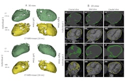
Using cardiovascular MRI, a team headed by Dr. Clara Hagedorn (who presented the findings) and Dr. Alexander Schulz from University Hospital Göttingen, Germany, sought to establish the connection of elevated epicardial adipose tissue (EAT) volume to myocardial infarction (MI) and the pathomechanism by which it contributes to adverse outcomes in MI.
EAT, the fat layer between the myocardium and the lining of the heart that directly surrounds coronary arteries, is already known to be associated with coronary artery disease and major cardiovascular events. Furthermore, MI mortality has been shown to be correlated with the extent of myocardial injury.
 Dr. Alexander SchulzEACVI 2025
To investigate the association of EAT volume (EATV) and the extent of myocardial injury after an MI, the researchers conducted a prospective multicenter study involving 1,168 patients. The patients underwent cardiovascular MR within 10 days after a percutaneous coronary intervention following an acute MI.
To analyze the results, the researchers divided the patients’ results into quartiles based on EATV.
All quartiles had higher percentages of male patients than female (Q1-Q4: 76%, 77%, 70%, 72%, respectively; p = 0.288). The patients in the quartile with the highest volume of EAT were older on average (Q1-Q4: age 63, 62, 62, and 66, respectively; p = 0.002) and had higher average body mass index scores (Q1-Q4: 27.4, 27.8, 28.0, 28.9 kg/m², respectively; p = 0.001).
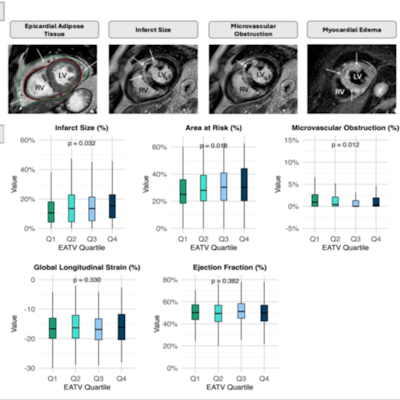
 Cardiac MRI analysis can provide useful information on epicardial adipose tissue, infarct size, microvascular obstruction, and myocardial edema.Hagedorn, Schulz, et al; EACVI 2025
While there were no differences in left ventricular ejection fraction among the quartiles, patients with higher EATV showed progressively increased infarct sizes (β = 0.064; 95% confidence interval [CI]: 0.005 to 0.122; p = 0.032) and larger areas at risk (p = 0.018).
However, those with higher EATV also exhibited less microvascular obstruction (β = 0.024; 95% CI: 0.042 to 0.005; p = 0.012). The authors note that no clear trend or pattern emerged from the data with microvascular obstruction, which demonstrated variability across the quartiles. As with left ventricular ejection fraction, global longitudinal strain remained comparable among the quartiles.
The larger infarct sizes and areas at risk exhibited by patients with higher EATV show greater acute myocardial injury, the researchers pointed out. “Higher EATV was independently associated with the extent of myocardial injury, suggesting a role in myocardial vulnerability and underscoring the potential relevance for personalized preventive strategies.”