VIENNA - Chest CT scans acquired at radiation doses as low as an x-ray showed lesions of all kinds conspicuously, using the latest dual-source CT scanner and iterative reconstruction. Individualized approaches tailored to specific lesion types cut the dose even further, researchers said on Friday at ECR 2017.
 Dr. Rishi Agrawal from Northwestern University.
Investigators from Northwestern University wanted to determine the minimum dose necessary to depict a wide variety of chest lesions. Measuring subjective as well as objective image quality, they found they could acquire all-purpose diagnostic images at doses of 0.1 mSv.
"Robust reduction of radiation dose for current chest CT examinations at levels similar to a chest radiograph is possible using low kV settings and [advanced modeled iterative reconstruction (ADMIRE)]," said study co-author Dr. Rishi Agrawal in his presentation.
How low can you go?
Dose reduction in chest CT has been the subject of significant research efforts, particularly for lung cancer screening and the detection of lung nodules at ultralow doses using iterative reconstruction, he said. Chest CT also benefits from natural high contrast between lung parenchyma and air, which allows for very low doses compared to other anatomic regions.
Even so, "more research is still needed on the effects of combining both the extremely low-dose parameters and the different image reconstruction algorithms to determine the conspicuity of different lung lesions," Agrawal said. "The question is, can extremely low-dose protocols be used to evaluate all lung lesions?"
The study aimed to find the lowest CT dose parameters for detecting lung pathologies in a phantom model. The group used a third-generation dual-source CT scanner (Somatom Force, Siemens Healthineers), the tube voltage ranged from 70 kV to 100 kV, and the tube current was fixed between 7 mAs and 75 mAs.
All images were acquired with ADMIRE at a collimation of 0.6 mm and slice thickness of 1.5 mm, with a Br59d kernel and iterative reconstruction strength of 3.
Two thoracic radiologists blinded to the CT protocol independently reviewed the randomized images on a PACS workstation, evaluating conspicuity on a five-point scale, where 5 was optimal with well-defined borders, 1 was indistinguishable from artifact, and 3 was distinguishable as a subtle lesion and not an image artifact. The mean score was accepted as the consensus score.
Objective image quality was measured as image noise (at the thoracic aorta) and contrast-to-noise ratio.
Ultralow for most findings
The minimum radiation dose with acceptable overall image quality was at 70 kVp and 10 mAs, the researchers found. The lesion conspicuity scores were similar between the two radiologist readers (p > 0.05), Agrawal said.
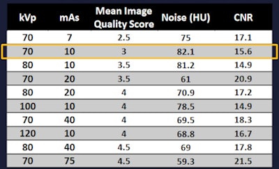 Results show minimum radiation dose with acceptable image quality for all lesion types. Images courtesy of Dr. Rishi Agrawal.
Acquisition settings that had a minimum score of 3 (representing subtle lesion and discernibly not artifact) for conspicuity varied according to lesion type. For example, bronchiectasis and ground-glass lesions could be distinguished at doses as low as 0.05 mSv, while the tree-in-bud finding required four times the dose.
It's "not surprising that with a solid nodule or bronchial polyps you could go right down to 0.05 mSv and still get a conspicuity score of 5," he said. "The only thing that was difficult to see was tree-in-bud opacity, which required an effective dose of 0.28 mSv to actually see well."
 Findings and minimum conspicuous dose levels by lesion type.
Limitations of the study included the use of a phantom without motion of any kind, and the use of only one scanner, Agrawal said.
"CT acquisition settings may be better tailored according to the clinical situation with very low doses for solid lung nodules and bronchial polyps, and robust reductions in radiation dose for chest CT exams at levels similar to a chest radiograph are possible," he concluded.


