Managers at a top U.K. hospital have made significant changes to how it uses abdominal radiography in the emergency department, following an extensive review of work patterns by a group of radiologists.
Abdominal radiography (AXR) constitutes an important part of the routine work-up of patients presenting with acute abdominal pain at most emergency departments, but an abnormal abdominal radiograph is notoriously difficult to interpret and a normal abdominal x-ray is misleading because it can miss various conditions. This is causing many hospitals to amend its procedures.
"Our experience from a busy university teaching hospital suggests that AXR often makes no significant difference in the management of these patients, who frequently go on to have further imaging with other modalities," noted Dr. Adam Yamamoto, from the department of radiology at Addenbrooke's Hospital in Cambridge, in a presentation at the recent RSNA congress. "Consideration must be made of a significant radiation dose (1 mSv), particularly in younger patients, but also the cost of unnecessary imaging and delayed time to final diagnosis."
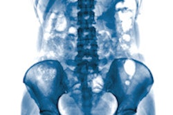
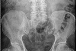
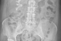
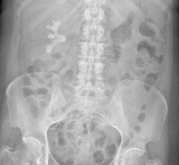 Digital abdominal x-ray shows large staghorn renal calculus, which is the white density below the ribs. But are such investigations of any great value when CT and ultrasound are more sensitive?
To evaluate the efficacy of AXR in the assessment of acute abdominal pain, and to provide evidence for a change of imaging practice, Yamamoto and colleagues conducted a retrospective analysis of abdominal x-rays performed for acute admissions to the emergency department and any subsequent imaging with CT or ultrasound. They included only patients who complained of abdominal pain, were aged older than 16, and were referred initially for AXR.
A total of 325 x-rays were studied. Of the 83% of radiographs that were normal, no further investigations were ordered for 51% of patients, while for the remaining 49%, CT only was performed in 37% of cases, ultrasound only in 19% of cases, and both CT and ultrasound in 7% of cases. For those undergoing CT only, 73% of the examinations were abnormal, compared with 50% for those undergoing ultrasound only or both CT and ultrasound.
For the 17% of radiographs that were abnormal, no further investigations were ordered in 38% of cases, while for the remaining 62%, CT only was ordered in half of cases, ultrasound only in 9% of cases, and both CT and ultrasound in 5% of cases. Of those undergoing CT only, 86% of examinations were abnormal, compared with 60% for those undergoing ultrasound only or both CT and ultrasound.
AXR is a standard clinical investigation, but is neither sensitive nor specific in the management of the acute abdomen, according to Yamamoto. Irrespective of the results of AXR, about half of patients will have further imaging with CT and ultrasound, in which a high percentage is usually abnormal. These imaging tests are performed on average over 24 hours after the initial presentation. During this time, patients leave the emergency department and are admitted to hospital wards, which has major cost implications for overnight admissions and places pressure on resources.
As a result of the study, the researchers recommended establishing an ultrasound service in the emergency department during daytime hours. Also, they advised clinicians to decide early if any imaging is required, and suggested that AXR should be avoided and the most appropriate imaging modality should be chosen. Ideally, these decisions should be taken by an experienced clinician.
Six months after making these recommendations, Yamamoto and his colleagues conducted a review of imaging practice. The outcome was a 216% increase in ultrasound examinations, plus a 33% decline in CT scans, and a 62% fall in AXR. Additionally, the average time from admission to further imaging with CT or ultrasound has fallen from more than 1.5 days to around 12 hours as a result of their suggestions.
"Abdominal radiography plays a limited role in the work up of patients with acute abdomen and should be replaced by other modalities. CT and ultrasound are more sensitive and more likely to provide an accurate diagnosis," concluded Yamamoto. "The risk is that abdominal radiography may simply be replaced by CT, and to this end, referral patterns and activity are being monitored."


