Dear Advanced Visualization Insider,
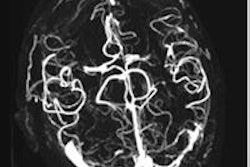
German researchers have developed a novel CT postprocessing algorithm to boost angiographic intensity in CT angiography of the head, spiking the contrast-to-noise ratio and improving visualization qualitatively as well.
Researchers from Ludwig-Maximilians-University in Munich developed the technique to aid visualization of the cerebral vasculature in acute ischemic stroke patients, finding they had increased contrast-to-noise ratio no less than ninefold. Learn how it works by clicking here.
In thoracic imaging, many people assume that computer-aided detection (CAD) picks up large lung nodules, say 30 mm in diameter, at least as well as smaller ones. But those people would be wrong. Structurally, the large ones are quite different and they require techniques for identification; CAD developers have been content to let radiologists pick up the large ones on their own. But in an era when CAD may be poised to take over the radiologist's first-reader responsibilities, this could be shortsighted. Check out a new large-nodule CAD scheme by Dutch researchers by clicking here.
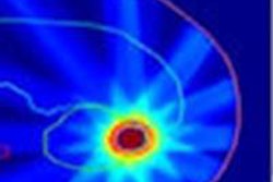
Radiology has been brought to you in living color for some time now. But medical imaging displays, still calibrated in grayscale, seem to have missed the memo. Change is afoot if a proposed extension to the DICOM Grayscale Standard Display Function is adopted. Enter the proposed Color Standard Display Function, and see how color calibration performs versus the old method by clicking here.
In radiotherapy planning, tumors move with the patient, shrinking and growing. Imaging is finally starting to keep up, according to presentations at a recent 4D treatment planning workshop in the U.K. For example, engineers from Utrecht in the Netherlands are aiming to use MR images as feedback for adapting treatment while the patient is on the table. Learn more here.
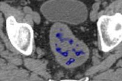
Finally, texture analysis is becoming increasingly important in oncologic imaging. Across many tumor types in several different organs, cancer's generally rough, heterogeneous patterns are offering clues to clinicians who want to avoid biopsies of benign lesions. The U.K.-based developers of Tex-Rad, the method behind a wide range of texture-analysis applications, have high hopes for its future as a breakthrough technology. Learn more here.
Be sure to visit your Advanced Visualization Community often to keep up with the latest developments. We also invite you to scroll through the links below for more news about the technologies that are making radiology diagnosis faster and more accurate for everyone.
