Dear Digital X-Ray Insider,
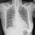
In chest examinations, digital tomosynthesis can offer improved diagnostic performance over conventional radiography because of the reduced visual clutter of overlying anatomy. The technique lacks the depth resolution of CT, but it provides high-resolution images in the coronal or sagittal plane with a substantial reduction in radiation dose compared with CT.
New research has compared digital tomosynthesis with digital radiography and CT in the thorax, and you can read more here.
Researchers from the U.K. have found radiographer-led immediate (or "hot") reporting in the emergency room can cut overall hospital costs, reduce treatment errors, and improve doctor confidence. Furthermore, if radiographer reporting roles were expanded to chest and abdomen radiographs, the total savings could be substantial, they believe. Get the story here, and check back Friday for more news about radiographer reporting.
William David Coolidge was an important x-ray pioneer, and this month’s history column focuses on his work and the immense contribution he made to medical imaging during the 20th century. Click here to learn more.
Increased use is being made of intravenous iodinated contrast media, predominantly in x-ray and CT examinations. It’s vital to be well-versed with the evidence-based literature to optimize image quality and minimize the risks to patients, particularly when dealing with pregnant women, according to new research. To read more, click here.
Lack of awareness of radiation exposure remains a major problem, especially among junior doctors, it seems. For the findings of a survey of members of the European Society of Residents in Urology, click here.
To find many more stories, go to the Digital X-Ray Community, and make sure you check back regularly.

