Postmortem CT is finding favor as an alternative to conventional autopsy alone. But Swiss researchers have added a new wrinkle -- postmortem CT angiography (CTA) -- which they found useful in detecting bone and vascular lesions during virtual autopsies, according to research published online on May 1 in Radiology.
 Dr. Silke Grabherr, PhD.
Bone lesions and vascular lesions are especially common in cases of traumatic death, such as falls from height, traffic accidents, ballistic trauma, and sharp trauma in homicides and suicides, lead author Dr. Silke Grabherr, PhD, told AuntMinnie.com. This makes postmortem CT angiography an excellent choice to investigate such cases, and it can be used independently or with autopsy.
"The possibility to view the vascular system in detail [with CT angiography] is a kind of a revolution in forensic medicine," she said. "For the first time, vascular diagnosis is possible similar to in clinical investigations. This allows us to detect causes of death that could not be found before."
Alternative to autopsy
The low maintenance cost and rapid examination of bodies with CT have led to its increasing use in postmortem diagnosis as an adjunct or replacement for conventional autopsy amidst recent declines in autopsy rates, the authors noted. Although postmortem CT is capable of facilitating diagnosis in most regions of the body, it offers limited visibility of vasculature, however.
"Visualization of the vascular system is of highest importance for legal and forensic medicine, since many causes of death are hidden in the vascular system," Grabherr said. "As the examination of the vascular system is still difficult during conventional autopsy and was impossible in 'virtual autopsy' by imaging, I had the wish to find a technique that could solve this problem and therefore advance postmortem examination techniques."
CT angiography might be able to help. In prior research, Grabherr and colleagues demonstrated the potential of using CT angiography instead of standard CT to improve the postmortem visualization of vascular lesions in a small sample population. Their protocol used a pressure-controlled perfusion device (Virtangio, Fumedica) to inject a mixture of paraffin oil and a contrast agent (Angiofil, Fumedica) into the femoral vasculature -- ultimately enabling the visualization of arteries and veins during CT angiography. They suggested that this enhanced imaging could help guide and optimize subsequent autopsy.
To expand the scope of their early work, they assessed the overall performance of this CT angiography protocol in the postmortem diagnosis and forensic analysis of 500 bodies from nine different European centers. Approximately 69% of the population was male, and the average age at death was 59.3 years. The most common reason for death was natural causes (52%), followed by localized trauma (28%), suspected medical error (11%), and polytrauma (9%).
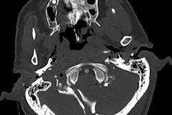
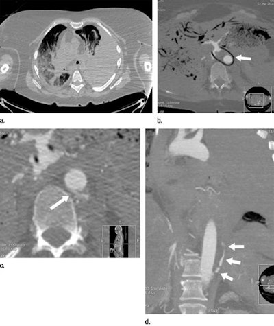 Postmortem CT (a) and postmortem CT angiography (b-d) scans of a 59-year-old woman who died of internal exsanguination soon after Whipple surgery. All images courtesy of RSNA.
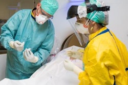
The group acquired CT and CT angiography scans of the bodies within five days of death. A forensic pathologist and a radiologist, both with more than five years of experience in reading postmortem CT angiography data, evaluated the scans individually. A second forensic pathologist conducted the autopsy for each case on the same or following day.
On their reports, the radiologist and forensic pathologists recorded 18,654 total findings, which they classified as essential (4,393), useful (9,540), or unimportant (4,721). They detected more findings with CT angiography than autopsy in all of these categories.
Best for trauma evaluation
Compared with autopsy, postmortem CT angiography revealed 5,347 (28.6%) more total findings and 601 (13.6%) more essential findings. Combining the two techniques led to the detection of more diagnostic findings than either technique alone or in tandem with standard CT.
| Autopsy vs. CT angiography for postmortem diagnosis | ||
| Autopsy | CT angiography | |
| Total findings | 61.3% | 89.9% |
| Essential findings | 76.6% | 90.2% |
| Skeletal lesions | 46.8% | 94.7% |
| Vascular lesions | 68.5% | 95% |
Postmortem CT angiography revealed a greater number of findings than conventional autopsy did, even when the cases were divided by type of wound. And this difference was especially pronounced for skeletal and vascular lesions, many of which were not even visible during autopsy.
The radiologist and forensic pathologists detected a statistically significant increase in essential bone and vascular findings using CT angiography compared with autopsy for all of the causes of death (p < 0.001). Specifically, CT angiography led to the discovery of 11.5% more essential vascular lesions in cases of natural death, 39.1% more in cases of localized trauma, 29.9% more in cases of suspected medical error, and 31.4% more in cases of polytrauma.
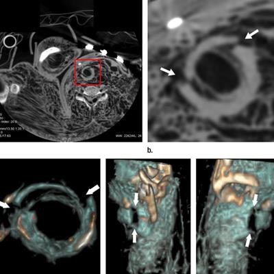
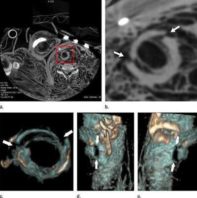 Cricoid cartilage of a 27-year-old woman who died of strangulation, visualized in an axial cervical postmortem CT scan (a, b) and volume-rendered 3D reconstructions (c-e). CT angiography revealed a bilateral fracture that proved difficult to demonstrate clearly with autopsy.
For some cases, the interpretation of the autopsy results concerning cause of death and the events leading up to it would have been incomplete or wrong without postmortem CT angiography, Grabherr said. This is why, in most centers, the forensic pathologist and radiologist work together.
"Whereas the pathologist is here to point out the importance of unfamiliar findings and explain postmortem changes in the body that can create specific artifacts not known by the clinical radiologist, the radiologist is here to see and read the images," she said. "Most literature recommends having a combined reading [by a pathologist and radiologist], especially as the field of forensic imaging is still young."
Future of forensic imaging
A key limitation of this prospective study was that, for legal and ethical purposes, the investigators were not able to blind forensic pathologists from receiving the imaging results before performing autopsies, the authors noted. It is possible that some of the findings might have otherwise gone undetected on isolated autopsy.
In the future, the researchers intend to add MR angiography to the mix of postmortem imaging modalities for the brain and liver, as they continue seeking out the best method to identify relevant findings when determining cause of death. They are also developing protocols for imaging infant bodies based on body weight, as well as testing various contrast agents that may be more suitable for scenarios involving advanced putrefaction or fatty embolism.


