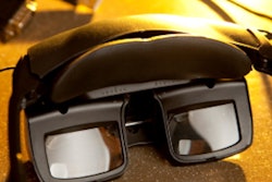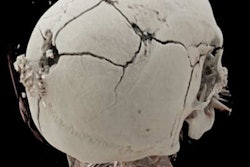
Imagine a jury able to observe a murder being committed using a virtual reality (VR) headset, and data taken from the victim's body. This isn't a scene from a futuristic sci-fi movie but a real-life experience for juries in Switzerland where radiologists and forensic experts are using postmortem imaging to recreate trauma mechanisms and crime scenes for the courtroom.
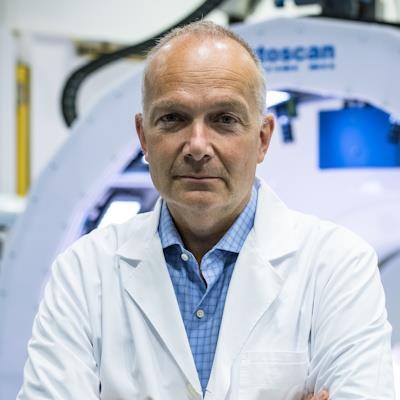
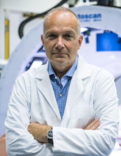 Dr. Michael Thali.
Two Swiss cases have used such technology. The first, now closed, involved a Swiss man who murdered his wife in the bathroom at home. The perpetrator who died some months ago while serving his prison sentence, had first attempted to murder his wife several years earlier in Mallorca by crushing her against a wall with his car as she came out of the house. The attempt failed and data captured by forensic CT imaging of the body was used to recreate details of the earlier attack in a reconstruction of events.
Speaking to AuntMinnieEurope.com ahead of ECR, Dr. Michael Thali, director of the University Zurich institute of forensic medicine, noted that after the car attack, the wife suffered from amnesia and the husband told the police that she had fallen from the first floor of the house. However, imaging and forensics told a different story, and the evidence helped to convict the man who was found to have killed his wife in order to claim a large life insurance policy. The other Swiss case using VR reconstruction is ongoing.
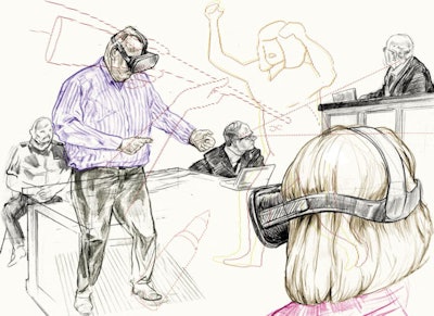 Virtually reconstructed crime scenes based on real 3D virtual autopsy data put defendants, lawyers, and judges at the scene of a crime with 3D glasses. Illustration courtesy of Christoph Fischer.
"This case clearly demonstrated that 3D reconstruction using postmortem imaging is worth it and not a novelty gimmick," noted Thali, who will be moderating a session on adult postmortem imaging on 2 March as part of ECR's "Radiology of the Afterlife" course taking place between 1 and 3 March.
Delegates at the session will hear about the latest techniques gaining ground in the "virtopsy" arena. Thali noted how in his institute MRI spectroscopy is being used to detect metabolic and functional changes in forensic cases, such as the level of alcohol in the heart or brain and other metabolic instability leading to death which can't be seen in a classical autopsy or on CT. The topic will be covered in detail by his colleague Dr. Dominic Gascho, in another Radiology of the Afterlife session on forensic imaging the next day.
"Postmortem MRI spectroscopy is still a niche area of ongoing research and not yet in daily use. However, I believe it will impact future postmortem scenarios because in standard autopsy and on CT you can't see or measure functional changes to detect a death from metabolic disorders such as diabetes. Spectroscopy can maybe even help to determine time of death in the future."
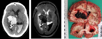 Brain hemorrhage due to hypertension or vessel anomalies is a natural death -- this finding in the postmortem imaging can allow such cases to be closed without open invasive autopsy. Images courtesy of Prof. Michael Thali.
CT triage
For Thali, the future of autopsy lies in imaging: In Zurich all bodies sent to the institute are scanned by CT which for the past five years, has served to triage cases for which the forensic questions can be answered through imaging, and cases which require classic autopsy in addition.
"Besides Melbourne, we are the only forensic institute doing this. If we can answer the forensic questions through CT or MRI, we don't cut up bodies. This speeds up workflow and saves money," Thali noted. "In Zurich we perform 500 classic autopsies per year, and up to 300 CT virtopsies. A classic autopsy costs two to three thousand Swiss Francs, two to three times higher than CT scanning which costs 900 Swiss Francs."
He described how a scan at 7 a.m. can lead to a decision to autopsy or not by 8 a.m. Visualizing an injury or pathology such as heart infarction or brain bleed, or foreign material such as bullets, and being able to determine whether a third party was involved or not meant that cases could be rapidly closed, not just money saving but also comforting for relatives.
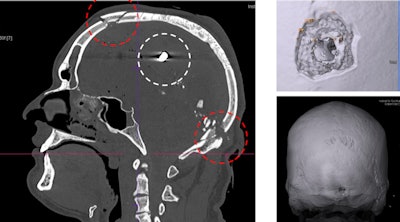 Gunshot injuries can be perfectly visualizised in 3D for court cases. Here, images show a gunshot with entrance wound in the neck area and bullet ricochet effect in the inner frontal part of the skull.
"Behind every body there is a family for whom we strive to answer questions without cutting up their loved ones," Thali said.
Of course, you can't see everything on images, he noted. If findings are unclear, then classic autopsy remains necessary. However, most forensically relevant and morphological questions can be seen by CT and MRI, with the possibility of postmortem CT angiography and image-guided biopsy.
Virtopsy access
In terms of postmortem scanners dedicated to autopsy, Switzerland is still ahead of the game as it was a decade ago, and this is largely in part due to the fact that many forensic doctors with radiological experience have become imaging department heads in universities across the country. This has meant the development of public forensic institutes boasting CT and some MRI scanners in Lausanne, Basel, Geneva, Berne and Zurich.
Elsewhere in Europe, access to virtopsy is unequal. Germany has, for example, an institute in Berlin that conducts postmortem scanning, while in the U.K., private religious organizations have some dedicated scanners but the only public forensic institute with dedicated postmortem imaging is in Leicester. Meanwhile, Thali's institute enjoys strong collaboration with the Victorian Institute of Forensic Medicine in Melbourne which sends its forensic medicine doctors to Zurich each March for a virtopsy course (see virtopsy.com).
Questions in the discussion may indeed cover this aspect of unequal access, with Swiss postmortem imaging taking place within the hour of a body's arrival when elsewhere living patients have to wait two to three weeks for their clinical MRI scans.
While such virtopsy initiatives are resource dependent, hospitals without their own dedicated forensic scanners should not be dissuaded from getting involved in postmortem imaging, according to Thali. The most important aspect is the connecting of forensic and imaging experts within departments and the chance to exchange and collaborate on projects. This way forensic doctors can gain access to imaging, and radiologists can learn about the forensic questions to be answered, he noted.
He pointed to an example of such collaboration: the creation by his team in 2011 of the multidisciplinary International Society of Forensic Radiology and Imaging (ISFRI.org) which meets annually -- this year in May in Toulouse -- and has its own journal "Forensic Imaging."


