All referring physicians must be made more aware of the current guidelines for requesting lumbar spine radiographs, as well as the legal responsibilities, to make certain the correct investigation is requested to answer the clinical question, U.K. researchers have advocated.
"We recommend circulating a document to GPs [general practitioners], radiology staff, outpatient departments, and other referral sources, summarizing the guidelines to ensure inappropriate referrals are rejected," noted Dr. Mariyah Selmi, a radiologist at the Royal Oldham Hospital in Manchester. "This will reduce the number of inappropriate scans, [cut] unnecessary radiation dose to patients, and ensure they are receiving the most appropriate investigation and treatment for their symptoms."
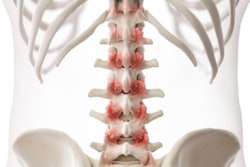
Nonspecific low back pain comprises tension, soreness, and/or stiffness in the lower back, for which it is not possible to identify a specific cause of pain. It affects about a third of adults in the U.K., and 20% of these patients seek advice from their GPs, Selmi told delegates at the 2016 U.K. Radiological Congress (UKRC) in Liverpool.
Under the definition of the U.K. National Institute of Clinical Excellence (NICE), lower back pain should exist for more than six weeks but less than a year, and there should be no red flags or alerts suggesting cauda equina or malignancy, and no evidence of trauma, for example.
Along with colleague Dr. Madhu Dutta, Selmi aimed to evaluate the appropriateness of lumbar spine radiography referrals for lower back pain. For the 762 lumbar spine radiographs performed in July 2015 by the Pennine Acute Hospitals National Health Service (NHS) Trust, they compared referrals with the 2009 NICE guidelines and the iRefer referral guidance from the Royal College of Radiologists.
The referral source was noted, and the following groups were broken out on the request card:
- Group 1: Lower back pain suggesting osteoporotic fracture (appropriate)
- Group 2: Nonspecific back pain (inappropriate, red flags/less than 6 weeks or more than 12 months)
- Group 3: Insufficient clinical details
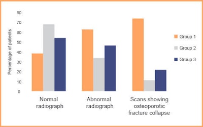 The graph demonstrates the percentage of images from each group that were abnormal and showed osteoporotic collapse or fracture. Most of the images from group 2 were normal. Source: Dr. Mariyah Selmi, presented at UKRC 2016.
About 29% of requests met the current guidelines, and of the inappropriate referrals, 62% had normal lumbar spine radiographs. Only 6% went on to show collapse or fracture. Of the abnormal radiographs in this group, 75% showed incidental abnormalities not thought to be in keeping with the request.
A total of 82% of referrals were from GPs.
Getting savvy on dose
Selmi and Dutta also think there needs to be a relationship between the referrer, practitioner, and operator on radiation dose awareness.
"The clinical details and reasoning given by the referrer will allow the practitioner and operator to justify the net benefit of the investigation and for all parties to fulfill their legal responsibility," they explained in a related study presented as an e-poster at ECR 2016. "Education of junior doctors and all staff needs to be expanded to impress the importance and serious implications of these aspects."
For the study, the duo conducted a survey that showed knowledge of radiation dose, responsibility of referrers, and risk to patient safety was extremely limited. The survey looked at junior doctors' knowledge of radiation doses for commonly requested investigations, estimation of adverse effects, and their knowledge of legislation and referral guidelines.
Of the 49 responses received, none correctly estimated the dose of abdominal x-rays, CT head or CT thorax scans, and abdomen and pelvis (CT TAP) scans (see figure). What's more, 43% underestimated the radiation dose of CT TAPs, despite 89% of responders routinely requesting this investigation.
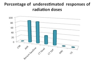 The graph shows the percentage of underestimates of radiation doses for routinely requested investigations. CXR = chest x-ray, AXR = abdominal x-ray, CT TAP = CT of thorax, abdomen, and pelvis, US = ultrasound. Source: Radiology department, Royal Oldham Hospital, Royal Oldham Hospital, Manchester, presented at ECR 2016.
A total of 27% of responders thought MRI and ultrasound used ionizing radiation, and only 49% and 20% correctly estimated the risk of inducing malignancy in CT TAP and chest x-ray, respectively. Of the 49 responses, only one doctor knew of any legislation and could name the Ionising Radiation (Medical Exposure) Regulations (IRMER) guidelines. In addition, none of the doctors surveyed had knowledge of the iRefer guidelines.
In the U.K., a number of pathways and protocols can be used to guide clinicians on imaging investigations. The most comprehensive, yet underutilized, is iRefer, Selmi and Dutta explained. This online pathway details symptoms and the most appropriate imaging technique to investigate them, as well as information on radiation dosing, and is an essential tool for doctors to ensure the correct investigation has been requested and should be accessed before each request, they wrote.
