Important changes in the epidemiology of congenital heart defects (CHD) are taking place, which mean these defects no longer concern only pediatric radiologists and pediatric cardiologists because an increasing number of cases occur within the adult population, according to new analysis from Austrian experts.
Improved treatment options, in combination with declining birth rates in western countries as well as more terminated pregnancies in severe malformations, mean that today there are more adults with CHD than children, noted Dr. Erich Sorantin, from the division of pediatric radiology in the Department of Radiology, and Dr. Bernd Heinzl, from the division of pediatric cardiology in the Department of Pediatric and Adolescent Medicine, both at Graz Medical University.
Echocardiography still represents the imaging modality of choice for morphological and functional assessment, and in childhood the different CHD types can be diagnosed confidently, and examinations can be performed at the bedside. In the follow-up of CHD, cross-sectional imaging plays an important role, and it is essential for radiologists to know the features, challenges, and limitations of echocardiography, they explained in an article-in-press published online by the European Journal of Radiology (EJR) on 29 May.
In the article, they present a systematic approach for morphological and functional assessment of the heart, along with representative images, and typical echocardiographic findings in common CHD are presented. In older children, adolescents and grown-ups with CHD, echocardiography suffers from limitations, partially due to skeletal deformations and lung emphysema. In particular, right ventricular function assessment is not always possible using echocardiography. Therefore, radiologist must be aware of the strengths and limitations of echocardiography and the role of cardiac MRI (cMRI) and cardiac CT (cCT), the authors stated.
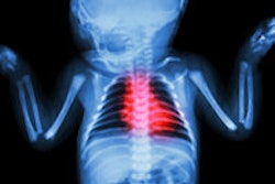
About 1% of all newborns suffer from CHD. Around half of cases consist of defects with shunts like the ventricular septum defect, atrial septum defect, and the persistent ductus arteriosus Botalli. The most frequent other types are (aortic) coarctation, pulmonary stenosis, aortic valve stenosis, and Tetralogy of Fallot. It is often possible to classify CHD according to the anatomy (obstruction of left or right heart, septal or vascular malformation, and complex CHD with anomalies of origin of the great arteries), or due to shunts (left to right, right to left, bidirectional shunt, or no shunt). Moreover, CHD types with and without cyanosis can be determined.
"Besides chest x-ray, echocardiography has become the most important noninvasive imaging modality of choice for evaluation of the cardiovascular system, providing morphological diagnosis as well as functional assessment," Sorantin and Heinzl wrote. "Beyond infancy, echocardiography is more difficult to perform due to restricted access, and cMRI provides a noninvasive, radiation-free imaging modality, which depicts cardiac morphology and function with high accuracy."
The purpose of their EJR article is to present the principles and limitations of echocardiography, diagnostic echocardiographic features of common CHD types, as well as the role of other imaging modalities. Though there are two major types of echocardiography, depending on the access (transthoracic and transesophageal echocardiography), they focus on the more common and basic transthoracic echocardiography.
"For transthoracic echocardiography there is no special preparation necessary. In rare cases, sedation (peroral, using chloral hydrate or midazolam) may be necessary in order to obtain diagnostic information. The patient is positioned on his left side down, in a reclined position in order to move their heart away from the sternum. For suprasternal views, the neck has to be overextended in order to place the transducer within the jugular fossa," they explained.
Sector transducers with a frequency range from 5 to 12 megahertz (MHz) are needed for proper near field view in premature/newborn infants, but for older children, 1 to 5 MHz devices are appropriate. Moreover, linear transducers are helpful especially in the assessment of the mediastinum and the supra-aortic vessels in neonates and infants. Besides 2D ultrasound, the machine should support M-Mode, pulsed wave Doppler sonography, color Doppler sonography, continuous wave Doppler sonography, and TEE. Further add-ons are 3D and 4D echocardiography, as well as tissue Doppler imaging.
"After basic patient evaluation (anamnesis, physical examination, ECG), echocardiography still is and will stay the first-line imaging method in childhood cardiovascular disease. In neonates, infants, and children, CHD types can be diagnosed by echocardiography, and functional heart evaluation can be done at the bed side, only right ventricular assessment is more challenging. In older children, adolescents, and adult patients, echocardiography suffers from limitations, where cMRI and cCT offer many options for complete morphological and functional heart assessment," the authors concluded.
