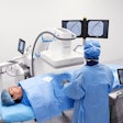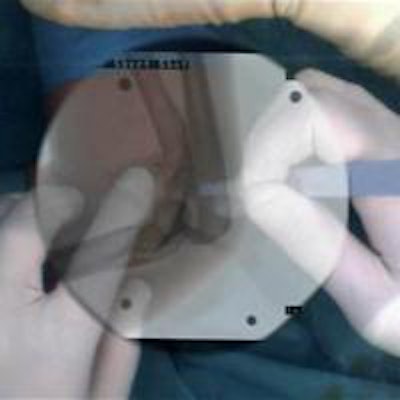
Accurate real-time depiction is key to appropriate diagnosis, treatment, and monitoring of patients when under challenging time pressures, and further advances in diagnostics will take place through intelligent systems allowing more meaningful mining of imaging and patient data, according to the president of the Computer Assisted Radiology and Surgery (CARS 2013) meeting, Dr. Nassir Navab.
Navab, chair of computer-aided medical procedures and augmented reality at the Informatics Institute at the Technical University of Munich (TUM), foresees a future in which surgeons and radiologists have access to crucial patient and image information at every moment of complex procedures. This area will be addressed at CARS 2013, to be held in Heidelberg, Germany, from 26 to 29 June.
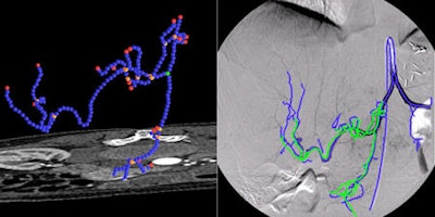 Complex catheter navigation for angiographic procedures can be supported by systems allowing the integration of preinterventional 3D image data into the intervention room. Via coregistration of pre- and intrainterventional images, physicians obtain more detailed information about current catheter positions and are able to follow predefined pathways through the vasculature. The first image (left) visualizes the 3D segmentation of vasculatures in CT angiography images used for planning a vascular intervention. The second image (right) visualizes the registration of the 3D segmented image used for diagnosis and planning and the 2D angiography, which is used for guidance during interventional procedures. All images courtesy of Dr. Nassir Navab.
"We are at the point of being able to create real patient and process specific imaging, of being able to predict and intelligently visualize what we need to see and interact with it; this flexibility is the real change and will greatly impact medical diagnostics and treatment," he said.
He also underlined the move toward knowledge-based medicine that allows decisions to be based on information extracted from data of tens of thousands of patients.
"Advances in diagnostics are less about new modalities and more about modeling processes from a clever use of available data," Navab said.
 With multiple disciplines at its heart, CARS brings together computer scientists, radiologists, surgeons, and industry figures to discuss the most advanced technology and its application in medicine. Delegates can hear firsthand how innovations impact healthcare, according to Dr. Nassir Navab.
With regard to computer-aided diagnosis (CAD), he said its current role as an "advisory" system particularly in breast and lung imaging has made -- and will continue to make -- the most impact in the screening scenario, despite its associated costs.
"As data systems become more comprehensive, CAD will be more useful for flagging potential diseases in patient populations. CAD's role will advance further and providers will have to make these systems more competitive price-wise," he noted. "There has been little resistance by radiologists to the introduction of CAD in the clinical setting; the specialist always has the final say in any decision-making even after using CAD technology."
Challenges to the integration of computer-assisted radiology and surgery in the clinical setting remain. There is still no intraoperative real-time multimodal and molecular imaging, which means it is not always easy for doctors to locate the region where intervention is targeted or immediately see the effects of their actions, according to Navab, who also leads Narvis, a research and development laboratory located next to the operating room units in two major Munich universities to which it is affiliated.
"Surgeons and interventional radiologists still rely on their eyes and personal knowledge, but in five to 10 years' time, intraoperative acquisition of molecular and functional images using modalities such as PET and SPECT will shorten operation time and greatly improve the accuracy of interventions," he said.
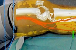
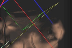
In addition, Navab pointed to the Narvis project CamC, currently used in the trauma unit of Klinikum Innenstadt at the Munich University Medical Center for orthopedic and trauma surgery. CamC is a conventional mobile C-arm equipped with a camera and a system of mirrors so that coregistration of the optical view and the x-ray image provides the surgeon with a "see-through" vision of bone beneath skin. This allows maximum intraoperative information at minimum radiation exposure to patients and medical staff.
So far, 40 patients have been treated using CamC technology, but it will be rolled out to three Munich hospitals in October for patient studies. Although mainly of interest to surgeons at present, further use of CamC for catheter placement may be envisaged in future.
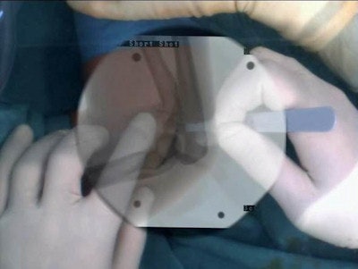 CamC enhances the conventional x-ray image with a coregistered video image. Physicians are able to switch between different views allowing optimal onsite planning and relocation of incision points.
Although changes to blood flow or overall blood circulation still cannot be computed in real-time, innovative research in procedures estimating and simulating blood flow using preoperative or interventional 2D and 3D angiographic imaging data will be outlined at CARS 2013.
"Flow simulations using computational fluid dynamics (CFD) techniques are already used by physicians to support treatment decisions. However, more research must be conducted in order to estimate and quantify blood flow during the interventions and predict changes induced by stents, flow diverters, artificial heart valves, or other vascular devices," Navab explained.
Also of note for interventional radiologists, is 2D/3D registration of angiographic images and catheter tracking for guidance of endovascular procedures. A series of studies using this technique will be presented by different groups who will describe their research in the navigation of catheters inside the vasculature for stent placement using a combination of preoperative CT angiography, intraoperative x-ray, and intravascular ultrasound.
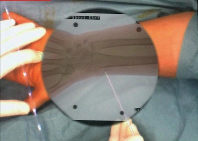 This image demonstrates the use of CamC system during a Kapandji procedure. The augmented reality view provided by the coregistered x-ray and optical imaging systems within CamC allows the surgeon to take one x-ray shot and execute the procedure under the guidance of the augmented reality view and in this way reduce the x-ray radiation dose in particular to the patient but also to the surgical crew.
"Such techniques allow doctors to make better decisions when faced with complications, through improving precision of stent placement, reducing the amount of x-ray radiation, and obviating the need for contrast injection," Navab said.
Other recent developments improving visualization and 4D physiological and functional understanding also will be showcased: Doppler echocardiography combined with angiographic imaging can provide a 4D model of the vascular system, which could, for example, enable doctors to check that artificial heart valves are functioning correctly.
A highlight of the congress will be a planned live transmission of a vascular intervention from the vascular surgery unit of Hospital Pasing, near Munich, led by Dr. Reza Ghotbi, whose team will use the most advanced angiographic techniques for minimally invasive stenting. A laparoscopic cholecystectomy procedure taking place at the Rechts der Isar Hospital in Munich under the direction of Dr. Hubertus Feussner also will be transmitted live. Both interventions will be discussed by a panel of radiologists and surgeons, who will describe the events in detail and debate what improvements are needed to improve workflow.
Research in minimally invasive robotic and interventional imaging will be presented on 26 June during the CARS satellite meeting, Information Processing in Computer Assisted Interventions (IPCAI). Here, experts in technology, surgery, and radiology will present their own most advanced research and describe new trend directions in multidisciplinary research.
"We would like to see more advanced computer technology inside radiology and surgery rooms. We need to take full advantage of the power of data analysis, machine learning, and deductive software technology," Navab said. "With advances being made in user interfaces and user interaction, we also need to bring standardization into the clinical setting -- can providers create solutions that function together?"
