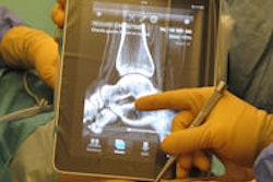
The iPad appears capable of supporting the diagnosis of pneumothorax on chest radiographs, according to an initial evaluation of an iOS-based image viewing app developed by a team from Massachusetts General Hospital (MGH). The group also found that its app, at least for now, didn't fare as well in cases with nonpneumothorax findings.
In a trial of MGH's RadPad app, chest radiograph interpretations in pneumothorax cases were highly concordant with those produced from a PACS workstation, but they were not concordant in exams with nonpneumothorax findings. Image quality and diagnostic confidence ratings were also high in pneumothorax cases but lower in those exams with nonpneumothorax results.
"Pneumothorax was detected better using the iPad as compared to infiltrates and effusions," said Dr. Supriya Gupta. She presented the research during a radiology informatics series on mobile computing devices at RSNA 2012 in Chicago.
Making rapid progress
Following U.S. Food and Drug Administration (FDA) clearance of the first iPad image viewing app in 2011, mobile-based image review has made rapid progress into radiology, Gupta said. As a result, the MGH researchers sought to assess the variations in diagnostic accuracy of the iPad with respect to radiological findings on chest radiographs.
Previous studies have validated the iPhone for urgent clinical diagnosis and for facilitating communication. However, the iPad, with its larger display screen and better screen resolution, offers the promise of improved speed and easier image navigation, she said.
As part of the institution's ongoing trial of the iPad, the team evaluated chest radiographs using its RadPad image viewer. RadPad is a native iOS app that is currently available inside MGH's institutional firewall network, with downloads and use restricted to specific users, Gupta said.
Over Wi-Fi, downloads take approximately 0.5 sec per image from an Amicas server, according to the team. Images are compressed at approximately 20:1, yielding a download speed of 250 msec per slice.
The researchers sought to assess image quality and diagnostic interpretation confidence based on certain radiological findings in an initial retrospective analysis of 30 radiographs using the iPad. The 30 chest radiographs were performed to exclude pneumothorax following tube placement in the intensive care unit (ICU).
The cases were first read in a blinded manner by a chest radiologist on the iPad; after one week, they were read again on a PACS workstation. The radiologist's various findings were recorded, and diagnostic confidence was graded on a scale of 1 (unacceptable image) to 5 (equal to PACS) for each category.
For pneumothorax-positive cases, image quality and radiologist confidence ranged from 4 to 5 with the iPad for the 15 cases. In addition, the observer findings were highly concordant with those produced from the PACS workstation (r = 0.9122, p = 0.0001).
It was a different outcome, however, in cases that included findings other than pneumothorax. In those 18 cases, image quality and radiologist confidence ranged from 3 to 4. In addition, observer findings on the iPad and PACS workstation did not correlate well (r = 0.64, p = 0.33).
These results led to a number of enhancements to RadPad after the study was performed, Gupta noted.
"[The RadPad] was not applying the value of interest from lookup tables in the DICOM header to transform the pixel data, so the contrast in the initial app was not as great," she said. "We have actually [now] made that enhancement and are building another trial for evaluating [its performance in nonpneumothorax cases]."


