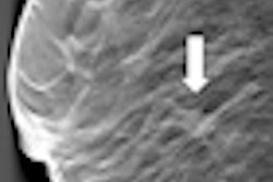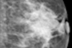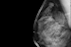
Researchers from Germany, France, and the University of California, Los Angeles (UCLA) have collaborated to produce ultrasharp 3D breast images with a technique that delivers less radiation than a mammogram, according to an article published online in the Proceedings of the National Academy of Sciences.
High-quality 3D breast tomography produces sharper breast images -- three times sharper than mammography -- that could enable the visualization of much smaller, earlier-stage breast tumors than are visible on standard dual-view digital mammography (PNAS, October 22, 2012).
Mammography provides only two images of the breast tissue, which explains why 10% to 20% of tumors are missed using the modality, said Dr. Jianwei Miao, a professor of physics and astronomy and researcher with UCLA's California NanoSystems Institute. CT scans can also produce 3D images, but they are rarely used for breast scans and would typically provide a much higher radiation dose than mammography, he said in a statement accompanying release of the study.
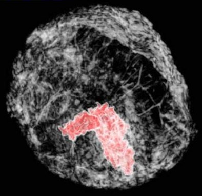 |
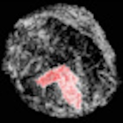
| The red area represents a 3D breast tumor. Image courtesy of UCLA. |
The UCLA researchers, along with colleagues from the European Synchrotron Radiation Facility in France and Germany's Ludwig Maximilian University, used phase-contrast tomography to x-ray a human breast from multiple angles. They then applied a new image-reconstruction algorithm, equally sloped tomography (EST), to the resulting images, using 512 images to produce very high-resolution 3D breast images.
In a subjective image quality evaluation by five Ludwig Maximilian University radiologists, the images were rated as having better sharpness, contrast, and overall image quality than 3D breast images created using other methods. The technique can also reduce radiation dose and acquisition time by about 74% compared with conventional phase-contrast x-ray tomography, while maintaining high image resolution and image contrast, the authors wrote in an abstract.
Unlike traditional mammography or x-ray exams that rely on differences in the intensity of the x-ray beam before and after it passes through body tissues, phase-contrast x-ray tomography measures the difference in the way an x-ray oscillates through normal tissue compared with denser tissues such as bone or tumor. Small tumors do not absorb x-rays well, but they substantially change the oscillation of the x-ray, Miao said. Phase-contrast tomography uses this oscillation difference to create the images, which are combined into the larger 3D image.
The primary contribution to the technique is the EST computational algorithm developed at UCLA, Miao said. A rethinking of the equations used in today's image-reconstruction algorithms led to the ultrasharp EST algorithm, which requires fewer image slices to produce a diagnostic image.
More research is needed before the algorithm is ready for routine use in patients. In particular, a high-quality x-ray source must come from a device small enough for breast cancer screening, said Alberto Bravin, PhD, managing physicist of the biomedical research laboratory at the European Synchrotron Radiation Facility.
"Once this hurdle is cleared, our research is poised to make a big impact on society," he said.


