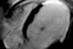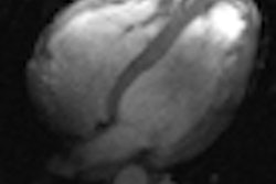
Cardiac MRI (CMR) finds significant numbers of extracardiac findings in patients with suspected heart disease, and as many as 20% are potentially serious, according to a Swiss study published online in European Radiology (4 January 2012). The prevalence of extracardiac findings has been investigated using CT but less with MRI.
MRI could potentially reveal more abnormalities due to its wider field-of-view, the authors said. In fact, the study revealed that about one-fifth of patients undergoing CMR were found to have potentially significant extracardiac findings requiring additional workup.
Interestingly, the second dedicated reading revealed significantly more extracardiac findings than the first reading, highlighting the need for training and for clinicians to pay more attention the first time around, the Swiss research team concluded. Few investigations have been undertaken of extracardiac findings in CMR, but their larger imaging volume may portend even more potentially serious findings compared with CT.
Cardiac MR "includes a substantial part of the thorax and upper abdomen, which may potentially contain noncardiac abnormalities that may be detectable incidentally on CMR examinations," wrote lead author Dr. Rolf Wyttenbach of Ospedale San Giovanni in Bellinzona, Switzerland, along with colleagues from University College in Bern and Spitalzentrum in Biel. "To our knowledge, no studies have evaluated the intraobserver agreement between the first clinical CMR reading and a dedicated second reading, where the focus was orientated to specifically detect extracardiac findings."
The study sought to determine the prevalence and importance of extracardiac findings in patients undergoing clinical CMR -- and to test the hypothesis that the original CMR reading focusing on the heart may underestimate extracardiac abnormalities.
The investigators scanned 401 consecutive patients (15 to 85 years, mean age was 53 ± 17, 66% men) who were referred for various indications, particularly ischemic heart disease (46%) and cardiomyopathy, myocarditis, or arrhythmia (together 41%). All patients underwent cardiac MR on a 1.5-tesla scanner (Intera, Release 11, Philips Healthcare) with 30 mT/m gradient strength and a 150 mT/m/msec slew rate, using vector ECG gating and a five-element phased-array cardiac coil.
Each exam followed a standardized protocol based on clinical indication, but all scans began with axial, coronal, and sagittal thoracic scout images with the following imaging parameters: balanced-turbo field echo; TR/TE 2.9/1.2 msec; flip angle of 50°; field-of-view of 450 mm; slice thickness of 5-10 mm.
Cine steady-state free-precession sequences (SSFP) in the standard cardiac imaging planes were included to assess systolic function, and depending on the indication, precontrast sequences were performed such as T1-weighted turbo spin-echo black-blood images, T2-weighted turbo spin-echo short tau inversion-recovery (STIR) black-blood sequences, and axial balanced turbo-field-echo (B-TFE) sequences during breath-hold covering the whole chest.
When indicated clinically, myocardial perfusion also was performed before and after intravenous adenosine infusion. And in all patients, assessment of late gadolinium enhancement (LGE) was performed using a 3D T1-TFE sequence acquired 10 to 20 minutes after 0.1-0.2 mmol/kg of gadolinium-DOTA (meglumine gadoterate, Dotarem, Guerbet).
In a dedicated second read for the study, the exams were read on a PACS (iSite, Philips Healthcare) for the presence of extracardiac abnormalities by a board-certified radiologist with 15 years of experience (R.W.) with fellowship training in cardiac MRI and expertise in body MRI. The reader, who had also performed the initial reading, was blinded to clinical data and results during the retrospective review.
Along with age, sex, and indications for the study, the report included the location and extent of cardiac findings, and the CMR sequences that best visualized it, categorized according to clinical significance.
"Potentially significant findings were defined as those that required further clinical or radiological follow-up, or that may have led to a change in patient management and should therefore have been reported to the clinician," the researchers wrote. "Insignificant findings were considered those that were more likely to be benign and not require follow-up." Follow-up was recommended for potentially significant findings that had not been previously reported.
The results showed a total of 250 extracardiac findings were detected, including 84 (34%) that were potentially significant including bronchial carcinoma (n = 1), lung consolidation (n = 7) and abdominal abnormalities. In 166 CMR studies (41%), nonsignificant extracardiac findings were logged. Almost twice as many extracardiac findings were documented on the second read (n = 84) versus the first (n = 47) by the same reader, and the number of findings identified as significant (84 versus 47 in the first read) and nonsignificant (166 versus 36 in the first read) was also significant (p < 0.00001), the authors reported.
Top potentially significant extracardiac findings (total n = 84)
|
The top findings deemed insignificant included kidney cysts (n = 57, 34%), liver cysts (n = 37, 22.3%), pleural effusions (n = 20, 12%), pectus excavatum (n = 12, 7.2%), and scoliosis (n = 5, 3%), Wyttenbach and colleagues reported. Only 32 (19%) were known before MRI, and 36 (22%) were detected during the first reading.
Of the 84 potentially significant findings, more than half (45/84, 54%) were previously unknown, and a little more than half (56%) were detected on the first read, which was focused on cardiac abnormalities. These included a necrotic pulmonary lesion in the lower left lobe of one patient which was a non-small cell lung cancer at diagnosis.
Recommendations for follow-up were made in 38 of the 84 (45%) patients with potentially significant findings, including five during the first reading and 33 during the second.
Among the 401 consecutive CMR patients, the prevalence of extracardiac abnormalities was 62% (250/401) with one-third (84/250) representing potentially significant findings, only 56% of which were detected at the first reading, Wyttenbach and colleagues wrote.
"Dedicated review of the CMR studies with special attention to [extracardiac findings] is therefore essential" they wrote, noting that the importance of such review is essential as emphasized by the detection of a previously unknown lung cancer. Moreover, missing significant extracardiac findings can delay proper therapy and could reduce the patient's life expectancy. As the use of CMR increases, the legal consequences and costs of missing such findings will probably rise as well, they said.
"While the clinical consequences of missing significant noncardiac lesions on a dedicated cardiac imaging study may be similar, there is, however, a major difference between CMR and [cardiac] CT: In CT, the whole cross-section is exposed to x-rays, and raw data for reconstructing it are available but usually not used; in MRI, only a circumscribed volume around the heart is investigated without the risk of ionizing radiation," Wyttenbach and colleagues wrote. "Thus, the ethical problem of missing significant findings is basically more important in [cardiac] CT than in CMR and may require whole-body as well as dedicated images."
"Our results show a significantly higher number of all [extracardiac findings], including the potentially significant ones, during the second imaging review with specific attention to noncardiac abnormalities," they wrote. "These findings highlight the importance of training and the specific review of the entire examination, including extracardiac anatomy in the chest and upper abdomen in order to detect potentially significant [extracardiac findings], which may affect patient treatment and outcome. Therefore, current cardiovascular MRI training guidelines should be adapted to include this topic in the curriculum of cardiovascular MR specialists, whether radiologists or cardiologists."
Potential limitations include the retrospective nature of the study and also the subjective definition of significant abnormalities.
About one-fifth of patients undergoing CMR studies have potentially significant extracardiac findings that may require further follow-up and may change patient management, they concluded.
The study "highlights the importance of a thorough look beyond the cardiac structures on CMR studies confirmed by the significantly higher number of [extracardiac findings] detected during the dedicated reading compared with the original clinical reading of the CMR study," Wyttenbach and colleagues concluded.


