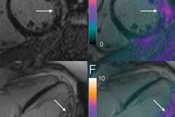
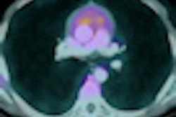
Dutch researchers have concluded that FDG-PET/CT can be a "sensitive tool," outperforming low-dose CT in determining the extent of bone involvement with sarcoidosis.
Researchers from Maastricht University Medical Centre in the Netherlands discovered a substantial number of sarcoidosis cases in the extremities using FDG-PET/CT, but a majority of those same lesions could not be detected by low-dose CT.
It's not known what causes sarcoidosis, which leads to inflammation in the extremities and can potentially affect any organ in the body. And it's also unclear how often bone involvement in sarcoidosis is present, said lead author Dr. Leonne Prompers, who presented study results at the Society of Nuclear Medicine (SNM) annual meeting last month.
"From previous literature, we think [incidence is] 2% to 3% of the patients, based mainly on morphologic or conventional imaging," she said. "The aim of our study was to examine the frequency and distribution pattern of bone involvement in sarcoidosis as detected by FDG-PET/CT."
The study included 123 patients who underwent FDG-PET/CT for severe sarcoidosis between June 2006 and September 2010, as well as a low-dose CT scan. All patients had unexplained disease-related disabling symptoms that persisted for at least one year and had sarcoidosis confirmed through biopsy.
The researchers excluded patients who suffered from other diseases that could cause a PET-positive finding. In the analysis of the 123 patients in the study sample, 94 (76%) had PET-positive findings associated with sarcoidosis. Subsequently, the 94 PET/CT scans were screened for the presence of bone or bone marrow localizations.
The researchers identified 32 patients (34%) with PET-positive bone or bone marrow localizations. Of those 32 patients, 19 (60%) also exhibited obvious focal bone lesions in a variety of locations.
"What is suggested in previous literature is bone lesions are predominately located in the extremities -- the fingers and the feet," Prompers noted. "What we saw was a lot of focal lesions in the whole skeleton, the axial skeleton, the pelvis, and the extremities."
Beyond extremities
The nonextremity lesions were found in the axial skeleton in 15 patients (47%), while 13 (40%) had lesions in their pelvis, 10 (34%) had lesions in their extremities, and one patient had a lesion in the skull.
Among 13 patients (40%), the researchers discovered large uptakes of FDG in both the axial and peripheral bone marrow, where no focal lesions were found. Both diffuse and focal uptake was seen in 11 patients (34%), while only focal lesions were detected in eight patients (25%).
"What is also quite interesting is that only two patients had lesions [detected] on their CT scans," Prompers added.
FDG-PET/CT-positive bone lesions were detected in one-third of patients in the study, Prompers and colleagues concluded. "That incidence is quite higher than expected, although this is somewhat of a selected population of patients who had severe complaints for more than one year," she added.
And while there was no single, most common location of the lesions, "I think we can say that PET is a potentially sensitive tool to detect bone marrow involvement," Prompers said.


