VIENNA - Innovation in software technologies, rather than shiny new hardware, provides the main talking points for visitors to the CT equipment manufacturers' booths at this year's ECR commercial exhibition. Companies are demonstrating novel approaches to reducing radiation dose, improving image quality, and extending the capabilities of this modality in imaging cardiac patients.
Iterative reconstruction software has become a well-recognized route to reducing patient dose in CT examinations, but Philips Healthcare is showing how this approach can also be used to produce virtually noise-free images and provide significant improvements in low-contrast resolution. Indeed, the company has suggested that the benefits offered by its new Iterative Model Reconstruction (IMR) process can provide a level of detail that is more commonly associated with MRI exams.
IMR has been made possible through the development of the company's first iterative reconstruction technique built on a knowledge-based model. The technology is enabled by innovations in both hardware and new algorithms that offer reconstruction speeds that allow IMR to be used in even the most demanding applications, according to Philips.
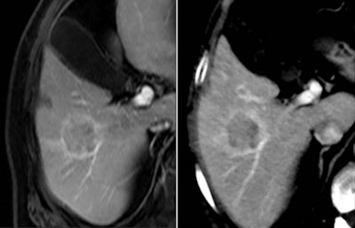 3-tesla MRI examination of the liver (left) compared with a CT image using the Iterative Model Reconstruction (IMR) technique. The lesion detected on CT was confirmed by MRI. Images courtesy of Philips Healthcare.
The main reported benefits of the new product are its low-contrast resolution with virtually noise-free images and a 2.7 times increase in low-contrast detectability. Philips warns that in clinical practice the degree of noise reduction will depend on the specific clinical task, patient size, anatomical location, and other factors. Therefore, a consultation with a radiologist and medical physicist is recommended to determine the appropriate dose to create the required image quality. However, the technology will typically offer less than five minute reconstructions for the majority of reference protocols, and is currently available with the Philips iCT and Elite class of scanners.
For its CT product range, the company is also offering two other ways of improving image quality: iDose and metal artifact reduction for orthopedic implants (O-Mar). The former is a means to improve image quality through increased spatial resolution and reduced artifacts at low dose. O-Mar reduces artifacts caused by large orthopedic implants. Available as part of its iDose Premium package, both features will become standard this year for 32-slice and higher members of the iCT and Ingenuity scanner families.
GE Healthcare is exhibiting a new scanner specifically designed to provide safe, high-performance scanning of patients with suspected cardiac conditions. The new Optima CT660 Freedom edition is available with intelligent coronary motion correction features and expanded capabilities for use in the emergency room.
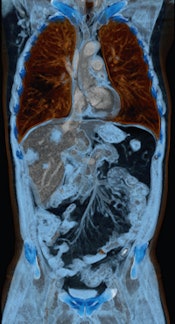 This case of a liver tumor was examined with a Somatom Perspective scanner from Siemens Healthcare. The volume-rendered technique (VRT) image highlights multiple liver lesions and fine details of the mesenteric arteries. Image courtesy of Diagnosezentrum Favoriten, Vienna.
Based on the company's FREE-dom technologies (Fast Registered Energies & ECG), the system offers a solution to "freeze" coronary motion in higher heart rate CT angiograms. This is achieved through the SnapShot Freeze component, which can significantly reduce coronary motion and overcome the inherent limitations of hardware-only solutions. By precisely detecting vessel motion and velocity, SnapShot Freeze can determine actual vessel position and intelligently correct the effects of motion during the exam, according to GE.
Users of the Optima CT660 system can scan quickly in one pass with diagnostic image quality and images reconstructed in real-time, so the scan will take only a few seconds, which may be vital in an emergency room setting. Meanwhile, in cranial studies of suspected stroke patients, the scanner also offers extended coverage of 80 mm with VolumeShuttle, providing twice the brain coverage for a single bolus of contrast at a lower dose than continuous acquisition techniques. The high-power capability and thin slices also provide the demanding clarity needed for detecting small lesions, the company said.
"We believe the traditional challenges of cardiac CT are in large part beatable with a smarter software and electronic approach," said Steve Gray, vice president and general manager of GE's CT business. "Freedom Edition offers physicians a new tool to help overcome coronary motion and various artifacts that may stand in the way of a highly accurate, confident cardiac diagnosis in a variety of clinical settings."
At its ECR booth, Hitachi is demonstrating an upgrade for the Scenaria 64-detector-row scanner that offers 128-slice reconstruction. The new Scenaria Advantage 128 platform features the company's second-generation iterative reconstruction algorithm, called Intelli IP (Advanced), which can double the speed of reconstruction from 18 images per second to 35.
After reducing the noise in the projection space based on an iterative process involving a high-precision statistical model, Intelli IP (Advanced) tunes image quality using anatomical and statistical information in the image space. This enables it to greatly reduce the amount of image noise and streak artifacts, the company explained.
Toshiba has unveiled a new version of its 320-detector-row scanner, Aquilion One, first launched in 2007. The latest version has been developed with a faster gantry rotation speed and a more powerful x-ray generator, giving clinicians the ability to perform cardiac imaging of patients with rapid heart rates in a single gantry rotation.
The Aquilion One Vision Edition is equipped with a gantry rotation of 0.275 sec, a 100-kw generator, and 320 detector rows (640 unique slices) covering 16 cm in a single rotation. With its 78-cm bore and fast rotation, the system can accommodate more patients, including bariatric patients and those with high heart rates.
The unit is equipped with the company's third-generation iterative dose reconstruction software, AIDR 3D, which can reduce radiation dose compared with conventional scanning.
"Aquilion One Vision Edition reduces risk and maximizes returns," said Satrajit Misra, senior director of the CT Business Unit at Toshiba. "It is capable of imaging the entire brain and heart in a single rotation with 500-micron accuracy, and can capture both anatomical and functional data."
The integrated AIDR 3D radiation dose reduction technology creates safer exams for improved patient care, he added.
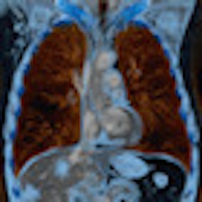
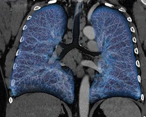 In this case of lymphoma, the VRT image shows multiple enlarged lymph nodes in the mediastinum and anatomical details in the lungs. Image courtesy of Clinique Sainte Marie, Paris.
Siemens Healthcare has announced a new member of its family of CT scanners: a 64-slice configuration of its 128-slice Somatom Perspective machine, designed to be an efficient and affordable technology. With a small footprint of just 18 square meters, an installation period of one to two days, and low energy consumption and cooling needs, the two Perspective scanners are among the most cost-effective systems in their class, according to the manufacturer. The new 64-slice machine is designed for use in outpatient environments and small to medium-sized hospitals where CT tasks focus on routine applications.
Both units are equipped with the eMode feature that automatically sets scanning parameters to run the equipment more efficiently, lowering maintenance costs and increasing the working life of the system.
Another feature of the new product is the FAST (fully assisting scanner technologies) CARE (combined applications to reduce exposure) platform, which aims to simplify and automate time-consuming and complex procedures before, during, and after the scan. This is intended to optimize the entire examination process, as it brings together all the steps involved, from scan preparation and performance through image reconstruction to diagnosis in a single integrated solution, Siemens claims.
The FAST CARE process has been developed to improve workflow, and the vendor is now offering it as standard for all machines in its range.
"Patients will benefit from an improved clinical workflow along with the greater availability of advanced clinical services like cardiac examinations. FAST CARE raises productivity, allowing medical professionals to spend more time with their patients," noted Jan Freund, head of CT product marketing management with Siemens Healthcare.
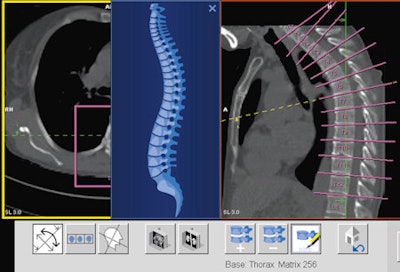 FAST Spine, a component of FAST CARE, can deliver an automatic segmentation of the spinal canal and automatic labeling of the vertebrae. Image courtesy of the University Hospital of Zurich.
Originally published in ECR Today on 8 March 2013.
Copyright © 2013 European Society of Radiology


