VIENNA - A declining birth rate and an increasing elderly population have resulted in new socioeconomic and healthcare problems. Physicians are confronted with complex clinical scenarios arising from this situation.
Diagnostic imaging plays a valuable role in the care of older patients. In the elderly, comorbidities compounded by physical and cognitive impairment can make it difficult for radiologists to provide the clinician with anticipated answers. It is important for radiologists to know the complex health scenarios occurring in geriatric patients, which are different from those seen in younger adults. They must be aware of the possibilities -- and limitations -- of imaging the geriatric population.
Sunday's European Congress of Radiology (ECR) Special Focus Session was designed to enhance doctors' recognition of the coexistence of various diseases in target patients, and distinguish the healthy older person from those in need of treatment.
 |
| Dr. Frederik Barkhof from Amsterdam. All images courtesy of the European Society of Radiology. |
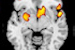
Key to management of diseases in the aging brain is understanding the structural visualized brain changes that occur in normal aging, which were presented by Dr. Frederik Barkhof, a professor of neuroradiology at the Free University of Amsterdam in the Netherlands. Widening in the Virchow-Robin spaces (VRS), as seen in MRI, is a normal aging phenomenon even in patients as young as 30, and of no pathological significance, he said. Some degree of atrophy of the medial temporal lobe was also sometimes visible.
Extreme widening in VRS was more difficult to determine. Patients often presented with headaches, but no discernible pathologies. It still might be a sign of normal aging, while homogenous dot-like structures of état Criblé was an abnormal finding indicating local atrophy often associated with diffuse white matter changes and always with a pathological finding.
"Normal aging is often defined as the absence of overt disease, but there are a lot of things happening below the threshold. There's a small group of people who, if you image them, have a healthy brain, but that's uncommon because most people with typical aging have some subclinical pathologies like white-matter lesions. The predictive value of these images is only partly known," Barkhof said. "Even if it's known, there are inferences at a group level, but on an individual level it is very difficult to make an exact determination of the impact down the line."
Determining whether patients will be able to live independently, and for how long, is crucial not only for the patients themselves, but also for their families. Unidentified white objects (UBOs) are probably a sign of mild hypoxia, and in a young subject should be reported as abnormal and lead to investigations. Once they start forming small bridges or become confluent, they should be considered abnormal, independent of age. Arterial spin labeling in subjects with extensive white-matter changes usually reveals poor perfusion of the tissue.
In one study, healthy subjects with severe confluent lesions (Fazekas grade 2 and 3) showed a 20% reduction in perfusion compared to patients with milder lesions. A LADIS study imaging patients with mild, moderate, or severe confluence showed that 60% of 639 subjects with severe white-matter lesions were unable to live independently at home after three years or had died, even though they were doing well at the time of scanning. The results indicate that healthy subjects with severe white-matter lesions have a poor prognosis, Barkhof pointed out.
Premature aging can be predicted through imaging markers, highlighting confluent white-matter lesions, silent infarcts, cerebral microbleeds, medial temporal atrophy, and amyloid markers.
ECR delegates heard how patients with Alzheimer's disease (AD) have increased amyloid binding, but presence of amyloid didn't necessarily mean a patient would develop AD or predict when they would develop it. However, functional biomarkers could show whether amyloid was interfering with brain function and also aid in predicting premature aging. New PET tracers could rule out AD in favor of other diseases.
"MR or CT should be used to work up suspected dementia and rule out treatable disorders, or suggest a specific diagnosis. If scans are negative, PET/SPECT should be used," he said. "PET/MRI can also help to diagnose early dementia, even short of established therapy."
 |
| Dr. Anne Cotten from Lille, France. |
Musculoskeletal disorders through trauma, degeneration, or malignancy were a particular problem for the elderly, delegates heard from Dr. Anne Cotten, head of musculoskeletal (MSK) radiology at Hôpital Roger Salengro in Lille, France. Loss of mobility and independence could have devastating effects on their quality of life.
Radiologists should know the most common MSK disorders as well as misleading presentations, potentially resulting in inappropriate or erroneous management. Besides low-impact falls from standing height, fractures caused by osteoporosis -- particularly vertebral fractures -- need careful attention as they are associated with excess mortality and significant morbidity, and because the risk of subsequent fractures was high, she said.
When vertebroplasty is indicated, MRI should be performed to localize vertebral fracture and stabilize the vertebroplasty, as well as deliver fast pain relief, Cotten continued.
It was also very important to be able to distinguish between osteoporotic vertebral collapse (VC) and metastatic VC. In osteoporotic VC, imaging findings showed multiple affected vertebrae in the thoracolumbar spine, with diffuse increased radiolucency and no osteolysis. Conversely, cases of metastatic VC were often unique, located in the cervical spine, with radiolucency and osteolysis. Missed vertebral fractures presented another pitfall for doctors.
"Rapid diagnosis is fundamental to avoid any delay in treatment, or the risk to the patient in this population is increased morbidity and mortality," Cotten said.
 |
| Dr. Tobias Saam from Munich. |
Partly focusing his talk on imaging features as potential predictors of cardiovascular disease, Dr. Tobias Saam, a radiologist at Ludwig Maximilian University in Munich, began by pointing out that this disease is a more common cause of death than cancer in any age group.
Age-related changes in vascular structure could indicate different stages of atherosclerotic development, and therefore risk factors of developing cardiovascular disease.
"Many imaging studies have shown that certain imaging features are associated with an increased risk; however, luminal stenosis alone is an insufficient marker to identify the vulnerable plaque," he commented.
Asked by session moderator Dr. Giuseppe Guglielmi, from the department of radiology at Foggia University Hospital in Italy, which modality was best to assess the risk factors, Saam responded that prospective studies were needed in PET/CT and MRI, both to determine the best methods to screen for atherosclerosis and identify the modality best suited to determine a patient's individual risk.
"We could be waiting five to 10 years for the results," he said.
Originally published in ECR Today March 7, 2011.
Copyright © 2011 European Society of Radiology


