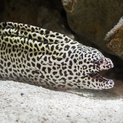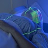
Even eels need medical imaging sometimes: Veterinarians at a zoo in Tacoma, Washington, used CT to image a lump that appeared in the mouth of a leopard moray eel named Larry Gordon, according to a report published on 28 October in the New York Times.
Veterinarians at Point Defiance Zoo & Aquarium noticed a lump in the 30-year-old, 16 lb (7.3 kg), 5-ft (1.5-m) long eel and initially determined it was probably due to a broken tooth, which was removed, according to a blog post posted by the zoo on 4 October. The lump disappeared, and staff thought the problem was resolved, but then the lump returned -- and was bigger.
"Because the growth returned, we decided to do a CT scan to evaluate Larry Gordon's delicate skull and dental features to ensure that removing the mass was possible and safe," Point Defiance veterinarian Dr. Kadie Anderson said.
Larry was taken from the zoo to a veterinary hospital in a water-filled cooler and anesthetized via an anesthetic added to the water, then placed in a smaller tub that fit the CT scanner. He underwent the scan, which revealed that the growth was isolated in the roof of his mouth and had not invaded his bone or nasal passages, the zoo said in its post. The team determined that it was not malignant and surgically removed it. Larry is recovering well, the zoo said.


