Dear Advanced Visualization Insider,
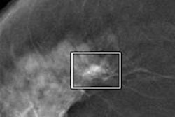
Mammography is the one of the few modalities that has fully embraced computer-aided detection (CAD) technology for daily clinical use, but CAD use is almost nonexistent in the fast-emerging area of digital breast tomosynthesis (DBT).
Now, researchers in Turin, Italy, are hot on the CAD trail with investigational CAD software that delivers high detection sensitivity with an acceptably low false-positive rate in DBT. The results were more than promising, but the study exposed a few wrinkles that show there's still plenty of work to do. Find out what's next by clicking here.
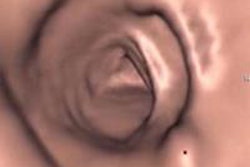
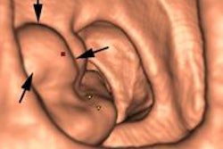
CAD is also pushing forward in CT colonography (CTC), where U.K. investigators are engaged in novel research to determine exactly what readers are looking at and what they're finding when they examine CTC data using CAD software. Certainly CAD helps readers focus on things they'd otherwise miss, but what about the elephants in the room? Could it be, for example, that by using CAD, readers are missing things they would otherwise find? Look for intriguing insights from a team that's been doing CTC and CAD since the early days, in an article you'll find here.
In Israel, Zebra Medical Vision is showing off its stripes as a different kind of image analysis company. In its goal to support the creation of higher-quality image analysis tools, it has built a database of 10 million imaging studies, against which prototype image analysis software can be tested. The goal, according to the firm's co-founder and CEO Elad Benjamin, is to identify risk earlier and hopefully save more patients' lives -- and maybe even a little money. Read the rest of the story by clicking here.
Segmentation of organs or specific anatomic regions is the first step for a wide variety of surgical procedures and interventions. But manual segmentation is quite time-consuming, a reality that spurred a drive for automated, or at least semiautomated, applications to speed segmentation processing. Many such applications have worked well. But the problem is that no matter how accurate, the schemes are all one-trick ponies. They do not work well in the setting of variability -- between scanners, between protocols, and, on occasion, even between patients.
Dutch researchers are applying a machine-learning process called transfer learning to segmentation of data in brain scans. Learn more about transfer-learning schemes and how they save time by clicking here.
Finally, we invite you to scroll through the links below to stay abreast of the many advanced imaging and visualization projects underway, all here in your Advanced Visualization Community.


