AI-driven systems significantly reduce radiation dose exposure for patients and improve diagnostic image quality in CT imaging, but the role of radiographers remains indispensable, an Italian-led study has found.
“CT radiographers’ clinical expertise and technical proficiency are critical for the proper implementation and monitoring of AI tools, ensuring their seamless integration into daily practice,” noted Cristian Colmo, lead specialist of the CT Dose Excellence Program and MRI-CT senior radiographer at Affidea, Padua, Veneto, Italy. “As AI continues to evolve, CT technologists serve as a vital bridge between cutting-edge technology and clinical application, safeguarding the accuracy and reliability of diagnostic outcomes.”
The main aim of the group’s study was to investigate how CT radiographers can use AI tools for patient autopositioning, scan length optimization, and image reconstruction tools.
Fifty patients between the ages of 31 and 81 underwent two thorax abdomen pelvis (TAP) CT exams over a period of 18 months as part of their oncological follow-up care. Both examinations were performed with contrast media injection and multiphase protocol. The first exam was performed with a traditional 64-slice CT scanner (GE Lightspeed SL or VCT 64-Slice) equipped with the first generation of iterative reconstruction (HIR, ASIR from GE Healthcare). The second exam was acquired using the newly installed AI-enhanced CT 64-slice (GE Revolution Ascend 64 SL) in the same imaging center.
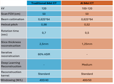 The CT protocol parameters used for both scanners. All figures courtesy of Cristian Colmo and presented at EuroSafe 2025.
All exams were conducted by the same team of radiographers, consisting of seven experienced members in the field of CT imaging, with experience ranging from 10 to 20 years.
The study, presented as a poster at EuroSafe 2025, used data for patient positioning in relation to the CT isocenter, scan range, and radiation dose indices to evaluate AI's effectiveness in improving imaging precision and reducing exposure. Radiation dose monitoring software (DoseWatch from GE Healthcare) was used to record and collect demographic, geometrical, technical, and dosimetric data. The team examined dose optimization by analyzing CTDI values of acquisition of the thorax, abdomen, and pelvis in the venous phase.
The radiographers’ performance was analyzed by measuring patient shift in the gantry compared with the isocenter and scanning length by comparing results with and without the use of two AI tools (Smart Plan and True Fidelity Deep Learning Image Reconstruction DLIR from GE Healthcare). Correct patient positioning in the gantry ensured accurate estimation of patient size based on scout images used for mA modulation. Overall image quality was assessed by an experienced radiologist with 25 years of experience.
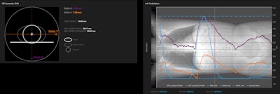 Detailed data on patient positioning in the gantry in relation to CT isocenter and mA modulation.
The results from 100 examinations were collected and analyzed using a business intelligence tool (Imaging Insights & Dose Analytics from GE Healthcare).
Radiation dose in terms of CTDI was reduced by 29% on average, Colmo and colleagues stated. A 40% reduction was obtained in obese patients, compared with 33% for overweight and 22% for standard-weight patients. Subjective image quality was evaluated as more than acceptable for traditional and AI-driven exams in all patients (respectively, 3.6 and 4.4 median values).
In regard to patient positioning and scanning length, AI-driven autopositioning delivered significant improvements in the accuracy and reproducibility, and in combination with smart planning tools, demonstrated a notable reduction in the time required for CT scan setup. The integration of these AI systems streamlined the workflow, minimizing manual adjustments and enhancing operational efficiency. Specifically, the AI algorithms accurately predicted optimal scan lengths, reducing the need for technologist intervention and ensuring consistent imaging protocols.
While the time savings were significant, no substantial differences were observed in the accuracy of the scan lengths as well as patient positioning when compared to traditional methods, the researchers pointed out.
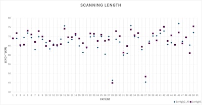 Scanning length for the two CT exams of each patient.
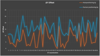 Vertical offset of the patient to CT isocenter for all examinations.
The group's results add support to the ongoing training of radiographers (as reported by DeWeese L et al, J Digit Imaging 2022 Apr;35(2):327-334). They also show the opportunity provided to each radiographer involved in the study to access the dose management software to analyze and review CT exam data, including patient centering and mA modulation, the authors wrote.
“These findings highlight the potential of AI to enhance workflow efficiency in radiology departments, allowing technologists to focus on other tasks while maintaining high standards of patient care. The integration of AI tools in CT imaging represents a transformative advancement in radiological practices, significantly enhancing both efficiency and safety,” they noted.
Overall, advances in AI are transforming CT imaging, highlighting the critical role of radiographers in implementing and leveraging such new technologies for optimizing patient care, managing radiation dose, and ensuring image quality, according to Colmo and colleagues.
“Their ongoing collaboration with radiologists and medical [physicists] will be essential to harness the full potential of AI, driving innovation while maintaining the highest standards of patient care. This synergy between human expertise and artificial intelligence promises to redefine the future of radiology, paving the way for more precise, personalized, and efficient diagnostic imaging,” they concluded.
You can access the full exhibit on the EPOS website of the ESR. The co-authors of the EuroSafe presentation were G. Schifano, A. Zompa, G. Zaggia, M. Codignola, S. Puggina, A. Papachristodoulou, A. Roncacci, C. Paraskevopoulou.


