French researchers are recommending against polyp surveillance for small colorectal lesions 6 to 9 mm in diameter, according to a new study in the August issue of Digestive and Liver Disease. The relatively high prevalence of advanced dysplasia in 6- to 9-mm lesions at colonoscopy renders the practice inadvisable, they said.
The prospective study performed at Conchin Port-Royal University Hospital in Paris found that more than one-third of polyps in the 6- to 9-mm category were advanced adenomas -- and so defined mostly due to the presence of a villous component, whose role in the development of cancer remains unclear. In addition, high-grade dysplasias were rare among small polyps, and no frank cancers were found in this size category (Digestive and Liver Disease, August 2011, Vol. 43:8, pp. 609-612).
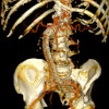
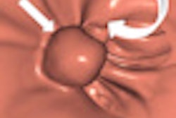
"Some authors have proposed that large polyps (> 10 mm) detected at CTC should trigger an immediate polypectomy, small polyps (6-9 mm) could either be referred for colonoscopy or undergo CTC [CT colonography, also known as virtual colonoscopy] surveillance, and diminutive polyps (1-5 mm) could be ignored," wrote Dr. Ulriikka Chaput and colleagues. "Many radiologists even suggest not reporting diminutive polyps because this size falls below the threshold for accurate detection with CTC."
The practice is controversial in polyps 10 mm and smaller, and the natural history of diminutive polyps 1 to 5 mm in diameter is not fully understood and needs more research, Chaput and colleagues added.
Among the few available studies on polyp surveillance, Hofsted et al performed annual colonoscopies but did not remove polyps smaller than 10 mm. Polyps 5 mm and smaller tended to grow, but polyps ranging from 6 to 10 mm in size tended to shrink over the three-year follow-up, suggesting that short-term surveillance of 6- to 9-mm lesions was potentially safe, Chaput and her team noted.
"On the other hand, amongst polyps < 10 mm, the rate of advanced adenoma is relatively frequent, from 6.6% to 12.5%, suggesting that removing these polyps could be preferable," they wrote.
The study aimed to evaluate the rate of advanced adenoma and high-grade dysplasia among small (6-9 mm) and diminutive (1-5 mm) polyps.
Between 2006 and 2008, the prospective study examined 1,468 individuals (53.1% male; mean age, 59.5 ± 14 years) using conventional optical colonoscopy. Of these, 323 patients (22%) were younger than 50 years of age and 250 (17%) were older than 74.
The main indications for colonoscopy in the study population included bleeding (11.1%), abdominal symptoms (21.5%), iron deficiency (1.7%), personal history of polyps (40.8%), and personal history of cancer (6.8%). Twenty-five percent of patients had a family history of colorectal cancer, and 15% did not have symptoms and were undergoing colorectal cancer screening.
Patients with a personal history of inflammatory bowel disease, Lynch syndrome, colonic polyposis, or infiltrating cancer were excluded from the study, and the authors prospectively collected information on conditions such as high blood pressure, diabetes mellitus, coronary artery disease, hypercholesterolemia, body mass index (BMI), and alcohol and tobacco consumption, as well as patients' personal and familial history of colorectal disease.
Standard (not high-definition) colonoscopies were performed using white light. Narrow band imaging and/or chromoendoscopy were options the endoscopists could choose to characterize a detected lesion. The endoscopists were able to choose the removal technique -- either cold snaring or biopsy forceps, Chaput and colleagues wrote.
For their analysis, the researchers distributed the polyps into two size groups: diminutive (1-5 mm, Group A) and small (6-9 mm, Group B). An open biopsy forceps was used as a 7-mm standard size. All pathological analysis was performed by two gastrointestinal expert pathologists working together, who classified the specimens by histological type (hyperplastic, adenoma, or serrated adenoma), degree of dysplasia, and presence or absence of a villous component (defined as 25% or more of the lesion).
The results in 1,468 patients showed 414 polyps smaller than 10 mm, of which 293 (70.8%) were adenomas, 41 (9.9%) were advanced adenomas, and 1.7% were high-grade neoplasia. Among the small (6-9 mm) polyps, 25 (35.2%) were advanced adenomas, mainly due to villous features, and three (4.2%) were high-grade neoplasia. Polyp size was associated with advanced adenomas (odds ratio = 8.47).
The researchers found no statistical differences in age, sex, high blood pressure, BMI, current smoking habits, alcohol consumption, or diabetes between the group with and without advanced adenomas. In fact, polyp size was the only identified risk factor.
"Considering polyps' characteristics, there was a global statistical difference between the morphological features of the two groups with more pedunculated (22.5%) and flat (10.0%) lesions in the advanced adenoma group, compared to the nonadvanced adenoma group (4.6% and 7.1%, respectively)," Chaput and her team noted.
There were more advanced adenomas in group B (35.2%) than in group A (4.7%) (p < 10-4). The diminutive polyps in group A were more often sessile (90.6%) than in group B (65.7%). There were also more tubulovillous adenomas in group B (32.4%) than in group A (4.1%).
"The rate of advanced adenomas amongst small polyps was 35%, mainly due to the presence of villous features," Chaput and colleagues wrote. "Polyp size was identified as a risk factor of advanced adenoma amongst polyps < 10 mm. Given these results, we believe that polypectomy should be warranted for patients presenting with small polyps at computed tomography colonography."
Risk factors associated with adenomatous polyps have been investigated, but data are lacking about risk factors of advanced adenoma, specifically among polyps 10 mm and smaller, which represent a larger percentage of detected polyps regardless of screening technique, they wrote. Older age has also been associated with advanced histology, but other studies did not reach this conclusion, they noted.
"A strong association was highlighted between polyp size (6-9 mm) and advanced histology within < 10 mm polyps," the authors wrote. "That observation offers a clear rationale to the 6-mm cutoff adopted for CTC."
The study found no major difference between advanced adenoma and high-grade neoplasia in diminutive polyps. However, there was a large difference between the rate of advanced adenoma (35.2%) and high-grade neoplasia (4.2%) in 6- to 9-mm polyps.
"That raises the question of the real importance of villous features as a risk factor of developing a colorectal cancer by itself," and it is still unclear whether the villous feature without high-grade dysplasia really increases the risk of colorectal cancer, the group wrote. Clear definitions regarding villous histology are lacking and reproducilibility is poor, they added, even though surveillance is based on polyp size and histology including the villous component.
In France, surveillance colonoscopy is at three years instead of five years in cases of polypectomy of a tubulovillous or a villous adenoma, instead of an exclusively tubular adenoma, for example.
"The rate of advanced adenoma and high grade neoplasia amongst diminutive polyps was low in our study, suggesting that diminutive (1-5 mm) polyps diagnosed by CTC need not be immediately removed," Chaput and her team concluded. "The rate of advanced adenoma amongst small (6-9 mm) polyps however was 35%, mainly because of the presence of villous features. Given these results, we feel that an approach of immediate polypectomy for patients presenting small polyps diagnosed by CTC is warranted."


