VIENNA - Ultrasound is famous for being a safe technique, but it can become perilous at times for the examiner, especially when the patient is a belligerent baboon or a crazy crocodile. The bizarre world of veterinary imaging was unveiled in a special lecture at the World Federation for Ultrasound in Medicine and Biology (WFUMB) congress on Friday evening.
Dr. Thomas Hildebrandt, a vet who specializes in reproduction management at Berlin's Leibniz Institute for Zoo and Wildlife Research (Das Institut für Zoo- und Wildtierforschung, IZW), explained to a packed audience how imaging animals can present both challenges and risks.
"When you work with wildlife, you are often challenged by physical obstacles, but it is also quite dangerous," he said.
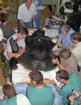 A male silverback gorilla was fully anesthetized by a team of human anesthesiologists at the Jersey Wildlife Preservation Trust in the Channel Islands. An ultrasound examination of the prostate and testes and also a semen evaluation were conducted to help ensure breeding success. A lesion on the gorilla's testicle was diagnosed using ultrasound, and it was treated with a hemicastration. All images courtesy of Dr. Thomas Hildebrandt.
Scanning pregnancy in specimens such as sea lions, sharks, and orcas exemplify both difficulties, but taking risks sometimes leads to interesting findings. When Hildebrandt and his colleagues were working in the Bahamas at the request of National Geographic, they discovered a new organ in sharks: the spiral valve. "This is a major part of the digestive track, truly an odd piece," he noted.
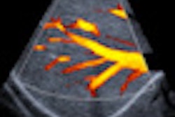
Vets may also encounter problems getting access to their patients. In his lecture, Hildebrandt showed a video featuring three Chinese doctors trying to grab hold of an uncooperative snub-nosed monkey, using only a small fishing net, for a color Doppler testicular examination and semen collection. He explained that IZW staff often work with monkeys, for instance in the optimization of ultrasound dosage protocols for the cloning of macaques.
Breeding can be problematic in captivity, and scientists are required for artificial inseminations. Sedation is vital to obtain the cooperation of an unwilling patient like a rhinoceros, but things can sometimes go wrong, and doctors must react quickly and decisively, he said. Another video clip featured Hildebrandt jumping repeatedly on a male rhino, which suddenly stopped breathing during the examination. The animal subsequently recovered and was responsible for two pregnancies within the next year, after having ignored his two female companions for 20 years.
"We don't know if that was the brain damage that made him like females better or if he was really afraid of anesthesia!" he quipped.
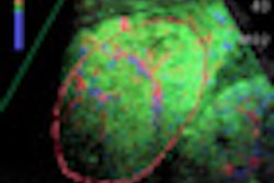
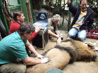 Male Asian lions are an extremely endangered species. This animal lived at Prague Zoo and was having breeding problems. His fertility was checked, including by means of an external ultrasound examination of the prostate and testes. After testing, it was found that the female had large ovarian cysts.
Among the other cases Hildebrandt has tackled are ultrasonographic sex determination of octopuses and komodo dragons, gynecological examinations of pachyderms such as elephants and hippopotami, performing transrectal ultrasound on a male hyena with prostate problems, and identifying the position of intra-abdominal testicles in Australian echidnas.
The size of some patients can pose a real challenge. At one extreme, IZW staff must scan the ovaries of a naked mole with an 80-MHz transducer. At the other extreme, they must determine the position of baby elephants in the uterus with a 2-MHz transducer.
Portable ultrasound devices are of great help in veterinary medicine, but users still struggle to provide high-resolution images and a good contrast level. To date, there is no image of a 15-day-old bovine embryo, for example. To obtain such an image, portable ultrasound devices with a resolution up to 100 micrometers would be needed; a resolution of which no mobile ultrasound device is currently capable.
"Since the 1980s, my research group has been in the lucky position to work with state-of-the-art equipment. We use ultrasound microscopes up to 80 MHz, 3D/4D high-performance sonographs, portable 3D/4D ultrasound devices, and the ultralight brand new V-scan for missions in Africa or Asia," he said.
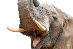
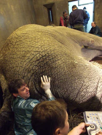 Fully grown anesthetized African elephant bull at the Zoo Dvur Králové in the Czech Republic. Hildebrandt and his colleagues performed a reproductive assessment using transrectal ultrasound, combined with an electro-ejaculation for semen evaluation. The testes were inside the abdominal cavity, located about 1.5 meters from the anus.
Hildebrandt would welcome the introduction of wireless connections between monitor, processor, and transducer, although he realizes this connection is likely to encounter practical problems. Portable 30-MHz devices also would be very useful additions to the range of veterinary ultrasound devices, but he still thinks veterinary ultrasound has a long way to go to before it reaches the same level as human use.
"In principle, classic veterinary medicine is about getting closer to the achievements of human medicine. So it's very important to use events like the WFUMB 2011 to show to a broad audience what ultrasound in veterinary medicine is capable of. Sometimes even experts of ultrasound in human medicine are quite surprised what we are able to achieve with ultrasound," he commented.
Hildebrandt has a special fondness for Vienna, particularly the Tiergarten Schönbrunn, which is the world's oldest zoo and is where the first test-tube elephant baby in Europe was born, and the first 3D imaging of an elephant baby was performed using a classic 530 Kretz machine. The zoo is one of Europe's role models for state-of-the-art animal keeping, he said.
Note: Additional reporting for this article was provided by David Zizka, a communications specialist in the European Society of Radiology's (ESR) press office.


