Superparamagnetic iron oxide (SPIO) is a tiny particle with huge potential to boost the detection of small lesions in the liver and prostate, speed up the imaging process, and do much more, according to a German radiologist.
Dr. Harald Ittrich from the department of diagnostic and interventional radiology and nuclear medicine at the University Medical Center Hamburg-Eppendorf (UKE) in Hamburg, elaborated on the growing clinical applications for these particles in an e-poster at RSNA 2017 in Chicago.
SPIO is often used in MRI and allows for imaging of anatomical, cellular, and molecular (pathological) changes. It also assists in interventional procedures, for instance in the liver, by looking at cell imaging, he noted.
SPIO summed up
SPIO particles measure 40 nm to 150 nm in size. They have fast phagocytosis by macrophages and a short blood half-life, and their primary indications include the liver, spleen, and bone marrow. In fact, SPIO can boost sensitivity of liver lesions to 97%, and for metastatic lymph nodes related to prostate cancer, SPIO can increases the sensitivity of native T2-weighted MRI from 35% to 91%, according to Ittrich.
SPIO also helps with magnetic cell labeling, tracking the movement of cells, and this can be helpful in the field of theranostics (therapy plus diagnostic). SPIO can diagnose cancer, target medication, home in on the tumor, kill the cancer cells, localize therapy, and improve imaging, he continued.
SPIO works in tandem with magnetic particle imaging (MPI). The magnetic fields move through a field-of-view, and SPIO registers a signal within the field free point. The signal correlates with the local SPIO concentration (approximately 30 nm). SPIO for MPI has "bright contrast," is quantifiable, with no nephrotoxicity, or feeding from the body's iron storage, Ittrich explained.
Two commercial examples include Resovist (Bayer Healthcare) and Feridex/Endorem (AMAG Pharmaceuticals). Resovist is an organ-specific MRI contrast agent, used for the detection and characterization of very small focal liver lesions. It consists of SPIO nanoparticles coated with carboxydextran, which are accumulated by phagocytosis in cells of the reticuloendothelial system of the liver. Uptake of the product in the reticuloendothelial cells leads to a reduction of the signal intensity of normal liver parenchyma on both T2- and T1-weighted images.
A comparison of MPI with other techniques is summarized in Ittrich's table below.
| MPI vs. established imaging modalities | |||||
| MRI | CT | PET/SPECT | MPI (recent) | MPI (theoretically) | |
| Spatial resolution | 100-200 µM | 200-300 µM | 1-2 mm | < 1 mm | < 100 µM |
| Temporal resolution (volume scan) | Sec./min | Sec. | Min. | Msec | Msec |
| Sensitivity | m-µMol | c-mMol | n-pMol | m-µMol | nMol |
| Penetration depth | 0 limitations | 0 limitations | 0 limitations | 0 limitations | 0 limitations |
Cousins to SPIO particles, ultrasmall superparamagnetic iron oxide (USPIO) particles, are 20 nm to 40 nm in size, primarily used in lymph nodes, he continued. As another difference, USPIO particles have a long blood half-life making them suitable for MR angiography.
Whether radiologists use SPIO or USPIO, these small particles have mighty potential, with more applications anticipated in the years to come, he concluded.
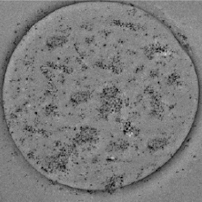
Editor's note: the image introducing this article on the home page shows a transmission electron microscopy image of an SPIO-containing microbubble. Courtesy of Dong Zhang, Nanjing University, China.


