A project led by the European Center for Research in Medical Imaging is developing a miniature PET/ultrasound probe to study new biomarkers for pancreatic and prostate cancer. Its aim is to create a bimodal imaging probe that combines a miniaturized time-of-flight PET detector head with an ultrasonic endoscope.
Furthermore, the PET section must deliver high sensitivity, a timing resolution of 200 picoseconds (ps) and millimeter (mm) spatial resolution.
The longer-term goal of Endo-TOFPET-US, which is now one year into its four-year duration, is to use the probe as a tool to develop new cancer biomarkers. At the International Conference on Translational Research in Radio-Oncology and Physics for Health in Europe (ICTR-PHE) meeting held earlier this month in Geneva, Paul Lecoq, PhD, senior physicist of the European Organization for Nuclear Research (CERN) in Geneva, told delegates about the latest progress in this international, multidisciplinary project.
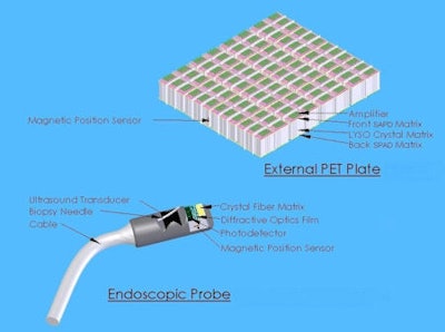
 The probe uses a split PET configuration, with one PET plate external to the body and the other within the endoscopic probe. Image courtesy of the European Organization for Nuclear Research (CERN).
The proposed probe design is based around a split PET configuration, in which one PET detector remains external to the body while the other resides on the endoscope tip, in front of the ultrasound transducer. The internal detector head can then be placed in near-contact with the region-of-interest.
This nonconventional design throws up a number of technical challenges. For starters, the asymmetry makes simulation and reconstruction a complex problem. The endoscopic design, meanwhile, requires miniaturization of the detector and associated electronics, as well as the ability to deal with constant motion and fluctuating, strong background signals.
To achieve the targeted millimeter spatial resolution, the internal PET detector head requires high granularity. To achieve this, the researchers created a matrix of scintillating crystal fibers, with each crystal measuring just 0.75 x 0.75 x 10 mm. The crystals are made from lutetium-yttrium oxyorthosilicate (LYSO) or cerium-calcium co-doped lutetium oxyorthosilicate (LSO).
The photodetector, meanwhile, comprises a silicon photomultiplier (SiPM) with a single-photon avalanche diode (SPAD) readout. This fully digital light detector enables single photon counting, thus maximizing the timing resolution. SiPM readout is performed using a low noise, front-end amplifier based on the NINO chip developed at CERN.
Lastly, a 500 µm thick diffractive optics film is coupled between the crystal matrix and the photodetector. This acts as a light concentrator to maximize the amount of light collected.
Site specific
The internal PET detector is coupled to a commercial ultrasound biopsy endoscope. The researchers came up with separate designs for use in the prostate and pancreas, the latter of which comes with more stringent anatomical constraints.
The detector head for prostate imaging, for example, is 23 mm in diameter and 22 mm long, and uses a matrix of 18 x 18 crystals (14 x 15 mm). The more compact pancreas design, meanwhile, is 15 mm in diameter and 22 mm long, with a 9 x 18 crystal matrix (7 x 15 mm). The prostate probe uses dual readout layers, while the pancreas probe uses a single readout.
The external PET plate measures 20.5 x 20.5 cm and comprises a matrix of 4,096 LYSO crystals (each measuring 3 x 3 x 15 mm) sandwiched between two SPAD matrices. During imaging, this plate will be placed on the patient's stomach. Clearly, to enable image reconstruction, the system must accurately determine the relative position of the internal and external PET detectors. To do this, an electromagnetic tracking sensor is placed on each one, enabling the system to reconstruct their positions in real time.
Lecoq presented some initial results from this novel PET design. In tests of crystal performance, an array of 16 of the smaller crystals exhibited a light yield of 10,300 photons/MeV and an energy resolution of 17%. The larger crystals produced similar results. "But the real innovation is the photodetector, we want to go to fully digital," he said.
Looking forward, the project team plans to launch a pilot clinical study focusing on pancreatic cancer, following preclinical feasibility tests on pigs. And will the device accomplish its target of 200 ps resolution? "Yes," Lecoq said. "Using our very low noise NINO amplifier we have already achieved better than 188 ps on the bench, with crystals of similar size and still analogue SiPM."
The European Center for Research in Medical Imaging (CERIMED) coordinates a European consortium of six academic institutions, three university hospitals, and three companies, and has received funding from the European Union Seventh Framework Programme (FP7/ 2007-2013) under Grant Agreement n°256984.
© IOP Publishing Limited. Republished with permission from medicalphysicsweb, a community website covering fundamental research and emerging technologies in medical imaging and radiation therapy.


