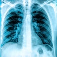
To help everybody who finds plain-film reporting challenging, award-winning Irish researchers have crafted a two-page checklist that can be used and referred to during the reporting process. The team won a certificate of merit at RSNA 2016 for its efforts.
The researchers, led by specialist registrar Dr. Stephen P. Power from Cork University Hospital in Cork, Republic of Ireland, tested out the efficacy of a printed checklist specifically for use in plain-film reporting and found all the junior residents reacted favorably to it and have incorporated the checklist into their early, plain x-ray reporting sessions. Specifically, it aids in the assessment of review areas and helps prevent "satisfaction of search."
"We acknowledge that the art of plain-film radiography cannot be condensed to two pages," Power and colleagues noted in their RSNA e-poster. "However, we think the checklist represents an accessible, although incomplete, learning tool for novice radiologists."
Checklist specifics
The checklist covers all areas of the body from head to toe by asking the reader questions of specific areas. For the shoulder, for instance, the checklist asks the following:
- Is the humeral head aligned with the glenoid process?
- Does the acromion align horizontally with the clavicle?
The checklist also has comments such as "assess for fracture" and "dislocation: 95% are anterior, 5% are posterior, often 2° to seizure/electrocution (ask yourself, is there a walking stick shape at the glenohumeral joint?)."
For the pediatric elbow, the checklist asks the following:
- Is there elevation of the anterior or posterior fat pads?
- Does the anterior humeral line pass through the middle of the capitellum?
For the adult elbow, the questions are similar to the pediatric elbow:
- Is there elevation of the anterior or posterior fat pads?
- Is the cortex of the radial head and neck smooth on both views?
Regarding the wrist, the checklist asks: Is the radial articular surface and ulnar styloid whole and intact? Is the scaphoid intact and normal?
For facial bones, is there fluid or soft tissue visible in the maxillary antrum?
In the cervical spine, assess first for adequacy (can you see from C1 to T1?).
For the thoracic and lumbar spine: Is there loss of height or wedging of vertebral body?
In the pelvis, assess the inner and outer contours of the main pelvic ring. Assess the two small rings forming the obturator foramina.
Regarding the knee joint, assess the patella and check for small osseous fragments.
The above represents a smattering of the material from the researchers. For the full list, check out Power and colleagues' e-poster.
They also noted that the checklist is a dynamic, evolving document that gets modified regularly based on resident feedback.


