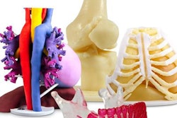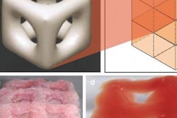
Scientists have developed a new microscopic imaging approach that improves 3D bioprinting and printed constructs, according to research published on 12 May in Bioprinting.
The research demonstrates the power of light-sheet fluorescence microscopy-based 3D real-time imaging to provide new insights into cell and fluid movements and flow patterns during extrusion using different bioinks, according to co-author Dr. Gowsihan Poologasundarampillai, a fellow in biomaterials and bioimaging at the University of Birmingham's School of Dentistry.
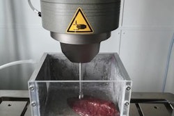
Additive manufacturing platforms create bioprinted structures by moving a special bioink that contains cells, biomolecules, and materials through a narrow tube, but the process can result in cells becoming damaged as they pass through the tiny tube.
The new technique illuminates a range of hydrogel-cell behavior and damage patterns depending on the extrusion speed and properties of the tube used during printing, according to a release from the university.


