A method that combines CT radiomics and real-time CT/ultrasound image fusion can yield more accurate and efficient tissue biopsies in ovarian cancer patients and may, in the future, even facilitate "virtual" biopsies, according to research published online in European Radiology.
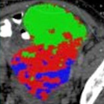
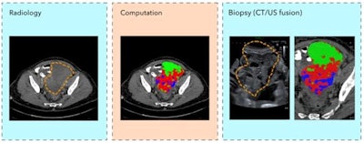
Researchers from the University of Cambridge in the U.K. implemented a targeted, ultrasound-guided tissue biopsy method that superimposed CT images with tumor habitat maps onto ultrasound images prior to performing targeted biopsies in patients with high-grade serous ovarian cancer.
In prospective testing on six patients, their technique provided a visual guide for physicians, aiding them to sample the full complexity of the tumor with fewer targeted biopsies, according to the authors.
"Our study is a step forward to noninvasively unravel tumor heterogeneity by using standard-of-care CT-based radiomic tumor habitats for ultrasound-guided targeted biopsies," said co-first author Dr. Lucian Beer from the University of Cambridge in a statement.
After performing tumor segmentation manually on prebiopsy contrast-enhanced CT scans, the researchers used spatial radiomic maps to identify tumor areas with similar or distinct radiomic patterns. CT images with superimposed tumor habitat maps were then co-registered with ultrasound data using Smart Fusion software (Canon Medical Systems).
Next, a gynecological radiologist with nine years of experience utilized the fused images to perform ultrasound-guided biopsy on all six participants using an ultrasound scanner with a 3.5-MHz convex transducer. Up to six biopsies -- two per tumor habitat -- were collected for each patient, according to the researchers.
 Individual and combined ovarian biopsy scans. Images courtesy of Prof. Evis Sala, PhD, of the University of Cambridge.
To determine the accuracy of the fusion method, the researchers used the dice similarity coefficient (DSC) to quantify the overlap of the tumor region between the ultrasound and CT images. The median DSC for tumor-specific CT/ultrasound was 0.53; higher accuracy was reported for larger pelvic tumors (DSC, 0.76-0.79) than for smaller omental metastases (DSC, 0.37-0.53).
The researchers noted, though, that the diversity of cancer cells within the tumor was successfully captured using this targeted biopsy technique.
"This will enable new approaches to discover and validate radiogenomic biomarkers," the authors concluded in the article posted on 14 December.
The researchers now plan to assess their method in a larger clinical study.


