Researchers from Israel have developed and tested a fiberoptic evanescent wave spectroscopy (FEWS) system that can noninvasively identify and characterize skin cancers. They successfully identified cancerous lesions on patients' skin by touching the suspicious regions for 30 seconds with an optical fiber connected to a mid-infrared (IR) spectrometer.
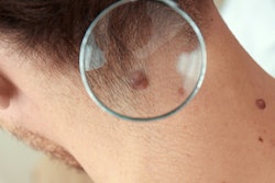
An estimated 300,000 cases of melanoma and more than one million cases of nonmelanoma skin cancer were diagnosed in 2018, according to the World Cancer Research Fund and the American Institute for Cancer Research. Suspicious lesions are usually identified by dermatologists using a dermascope, a handheld optical magnifier, and are subsequently diagnosed based on pathological analysis of biopsied tissue. This process, however, is invasive, costly, time-consuming, and dependent on the skill of the physician.
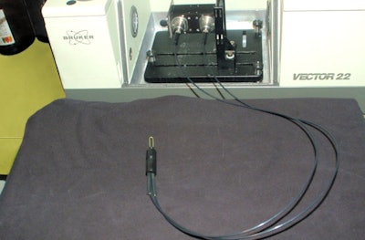 The sensor system is based on a spectrometer and a mid-IR transmitting fiber. When the exposed central part of the fiber (the U-shape) touches a skin lesion, the system measures its characteristic mid-IR absorption spectrum. Image courtesy of Abraham Katzir, Applied Physics Group, and Tel Aviv University.
To address these shortfalls, a group of researchers led by Prof. Abraham Katzir from Tel Aviv University is working to create an accurate, affordable clinical system that can identify skin cancers in near-real-time and could be used by dermatologists for reliable screening of suspicious lesions. Writing in Medical Physics, the researchers described their achievements to date.
The FEWS system is based on a mid-IR spectrometer and a long, U-shaped, mid-IR transmitting AgCIBr fiber. The fiber, developed by Tel Aviv University's applied physics group, is flexible, nontoxic, nonhygroscopic, and highly transparent in the mid-IR. The system operates in the 3-30 µm spectral range, to measure the mid-IR absorption spectra of tissues. To record an absorption spectrum, the center of the U-shaped fiber is simply brought into contact with the skin.
Patient studies
The researchers aimed to use the FEWS system to detect and identify three skin cancers: melanoma, basal cell carcinoma (BCC), and squamous cell carcinoma (SCC). For 90 patients in the dermatology department of the Tel Aviv Sourasky Medical Center, the authors measured the absorption spectra of each suspicious skin lesion and the surrounding healthy tissue, a process that took about 30 seconds per measurement.
The absorption spectra of the measured samples exhibited several peaks in the mid-IR region. Unique peaks at similar wavenumbers and within specific spectral ranges were easily identified by the naked eye as signatures of melanoma, BCC, or SCC. The team created "biochemical fingerprints" for samples of melanoma, BCC, SCC, and healthy tissue, based on differences in the absorption spectra. These spectra corresponded with the pathology of the patients' tissue biopsies, which included five melanomas, seven BCCs, and three SCCs.
In a recent refinement of their diagnostic method, the researchers developed an algorithm to analyze the spectra and deliver the results. For melanoma, this algorithm achieved 100% sensitivity, specificity, and accuracy.
One potential obstacle with this approach is that mid-IR radiation only penetrates only a few microns into the stratum corneum, the upper layer of the skin. The researchers suggest that this thin layer would include cells that have migrated upward from deeper malignant lesions.
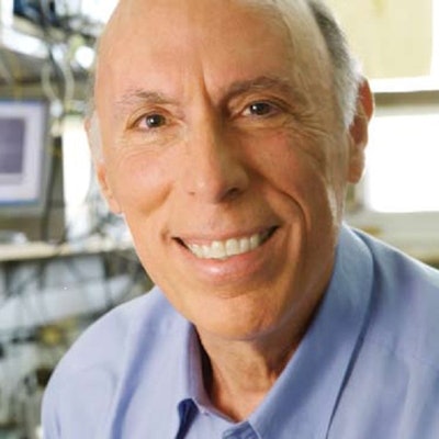
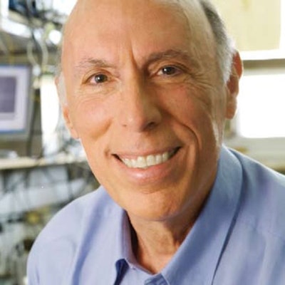 Prof. Abraham Katzir.
"This study confirmed that the melanoma-affected cells migrate from the layers of the skin to the thin top layer," Katzir explained. "We were able to use mid-IR spectroscopy on this very thin layer of skin to detect these cells, without breaking the skin, and from that to determine the type of cancer present. The differences among the cancer types were large and were immediately noticed by simple comparison of the spectra. This noninvasive 'spectroscopic pathology' may, in the future, replace the standard invasive biopsy."
He said the team is planning to conduct a larger number of experiments in a collaboration with the Sheba Medical Center, which has a large medical clinic dedicated to skin cancer.
"Our main interest is to develop a system that will automatically diagnose lesions, independent of the skill of the physician," Katzir added. "It will determine if the lesions are cancerous, and conclude whether they are melanoma or less-lethal cancers, all in real-time and in the clinic. The dermatologist community and the medical authorities are looking for a method that will diagnose, with a success rate of more than 95%, both malignant and benign skin lesions. If we succeed, we will try to get an approval for the method and then we will be ready for commercialization."
The next step will be to collect more data to improve the data analysis. The system also needs to be refined. The researchers plan to replace the commercial mid-IR spectrometer with a quantum cascade laser (QCL)-based system that will only cover the spectral range of interest, have higher intensity, and be more compact. They also plan to improve their software algorithms to better detect melanoma and other pathologies and to generate these findings automatically.
"We hope to develop a system that is small, lightweight, rugged, very easy to operate and, most importantly, inexpensive," Katzir said. "We want to reduce the cost to be less than 4,000 U.K. pounds (4,400 euros) so that small clinics can afford to purchase it. This system has the potential to make a sea change in the diagnosis of skin cancers and possibly other types of cancer. This is our goal."
Cynthia E. Keen is a freelance journalist specializing in medicine and healthcare-related innovations.


