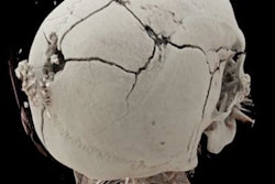
Once referred to as "3D on steroids," cinematic rendering (CR) has now become a powerful and effective tool to present pathologies found in postmortem CT for judicial reviews. That's the view of German researchers who have evaluated over 100 fractures and soft-tissue injuries.
"When we first tested cinematic rendering in 2017, we immediately saw the huge difference between it and the classic volume rendering (VR) technique, with CR creating more vivid, photorealistic reconstructions," Judith Böven, scientific associate at the department of diagnostic and interventional radiology, Düsseldorf University Hospital, told AuntMinnieEurope.com.
 Rib fractures with substantial dislocation of the third rib (green circles). Left to right: CT given a Likert score of 2 (good expressiveness, good visualization of the finding), CR given a Likert score of 1 (high expressiveness, very good visualization of the finding), VR with a Likert score of 2 (good expressiveness). All images courtesy of Judith Böven and the BJR.
To validate this impression and to discover if CR is superior to VR and classic 2D postmortem CT, the group performed a study in collaboration with forensic pathologists who are experienced with judicial reports. They published their results in an online article published on 18 June by the British Journal of Radiology, which is celebrating its 125th anniversary in 2020.
The key points
Between February 2016 and March 2019, Böven and her colleagues analyzed 112 fractures and injuries from 33 human cadavers (8 women, 25 men, with a mean age of 54 ± 18 years, range 31-92 years) that underwent whole-body postmortem CT after traumatic death. They reconstructed pathologies with CR and VR techniques, and classified fractures according to their dislocation.
Two forensic pathologists evaluated images according to their expressiveness and judicial relevance, and they decided whether CR reconstructions were suitable for judicial reviews. Two radiologists determined the detection rate of pathologies, and a traumatic cause of death was included.
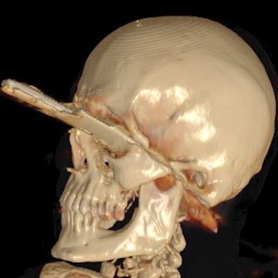
CR was more expressive than VR for all three trauma categories (p < 0.01). Also, CR was more expressive than conventional CT when used for fractures with dislocation (p < 0.001), injuries of the ventral body surface (p < 0.001), and demonstration of foreign bodies (p = 0.033). CR and VR became more expressive with a higher grade of fracture dislocation (p < 0.001). In total, 20% of all pathologies in the CR and VR reconstructions were not detectable by radiologists.
 Remaining knife. Left to right: CT image with a Likert score of 3 (medium expressiveness, finding can be detected), CR with a Likert score of 1 (high expressiveness, very good visualization of the finding), VR with a Likert score of 2 (good expressiveness).
"CT showed the same evaluation for expressiveness given by forensic pathologists in every grade of dislocation, whereas a strong correlation between grade of dislocation for fractures and CR expressiveness was found, as well as for VR reconstruction images," the authors wrote. "Reconstructions of fractures with major dislocation were declared useful for forensic reports in 84% of the cases."
In cases of fractures of smaller bones like the orbita or midface, forensic pathologists often found the CR and VR reconstructions not useful. A possible explanation could be lower digital resolution for smaller bones embedded in soft tissue.
 Blowout fracture of left orbita (blue arrow). Left to right: CT reconstruction with a Likert score of 1 (high expressiveness, very good visualization of the finding), CR with a Likert score of 4 (lower expressiveness, finding can hardly be detected), VR with a Likert score of 5 (very low expressiveness, finding cannot be detected).
"Due to the findings of our study, the use of CR for judicial reviews but also for other clinical demonstrations became the method of choice in our institute," Böven pointed out. "We also use CR reconstructions to demonstrate soft tissue injuries like stab wounds and penetration of foreign bodies more often than before."
The team's main conclusions were the following:
- CR reconstructions are superior to VR reconstructions regarding the expressiveness in all three trauma categories.
- CR was more useful than conventional postmortem CT slices in cases of fractures with substantial dislocation, demonstration of foreign bodies, and body surface injuries not covered by other body parts or the CT table.
- CR cannot be used for diagnostic purposes without postmortem CT, especially for fractures without substantial dislocation and covered soft-tissue injuries.
- CR can improve the interdisciplinary information transfer and the comprehension of forensic findings.
"The number of patients undergoing forensic imaging in our institute has risen in the last few years. This has led to a lot of interesting ideas for further studies in our research group besides creating 3D reconstructions," she stated. "For the current study, we excluded patients under 18 years old, so we definitely want to take a closer look at those patients offering new study areas."
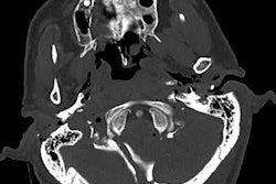
 Fracture of the os hyoideum. Left to right: CT image, CR image, VR image.
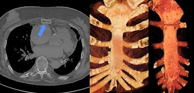
 Fracture of the sternum. The fracture cannot be detected on either CR or VR because of the missing dislocation. Left to right: CT image, CR image, VR image.


