A research group from a leading London facility won Europe's solitary magna cum laude for its research into the strategies, applications, and limitations of cinematic rendering of musculoskeletal MRI. The RSNA judges gave out just 23 of the prestigious prizes to the authors of nearly 2,500 digital posters displayed at the virtual congress.
"Cinematic rendering improves communication between radiologists, patients, and clinicians and enhances the sensitivity and specificity of radiological imaging, improving decision-making and facilitating patient-tailored management planning," explained Dr. Danoob Dalili, a radiologist at the School of Biomedical Sciences at Kings College London, and colleagues. "[Cinematic rendering] paves the way for integration of more advanced multidimensional visualization technologies including virtual and augmented reality."
Cinematic rendering comprises state-of-the-art postprocessing for MRI, providing far superior resolution and image interrogation to all previous forms of volume rendering (VR), and it is an example of radiologists working closely with industry and other disciplines to incorporate lessons learned in other parallel professions and recruit them to improve patient care, they noted.
Comparison with other methods
VR techniques display a 2D or 3D projection of images of large datasets, and they are particularly successful in interrogating spinal acquisition of CT due to the inclusion of complete volumetric datasets (no slice gaps). Studies show that 3D VR techniques have superior clinical applicability and accuracy when compared with maximum intensity projection (MIP) and shaded surface display (SSD) techniques, and this has not been surpassed over the years, according to the authors.


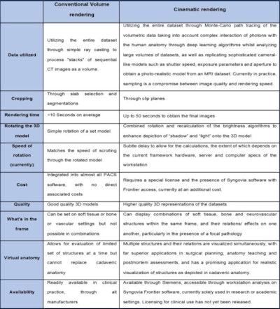
Key differences between cinematic rendering and convention volume rendering. Applying conventional VRT or cinematic rendering to MRI enables simultaneous visualization of all anatomical structures within a body part, and better understanding of the interrelationships between those structures. Table courtesy of RSNA 2020 and Dr. Danoob Dalili.
VR is less susceptible to artifacts, resulting from manual manipulation of data required with other methods, and unlike MIP, it can accurately illustrate two or more tissues adjacent to each other that possess different tissue characteristics – for example, grey and white matter in the brain -- as well as assess the data available within the entire dataset rather than a selected segment.
Novel applications
"Appreciating 3-D anatomy and the extensive pathologies within and around the spine is a key component of preoperative planning. [Cinematic rendering] provides 3-D templating to enhance knowledge and understanding of the surgeons, augmenting their choices of surgical approaches," they noted.
A recent preclinical study based on 720 evaluations illustrated that cinematic rendering visualization was associated with faster and more accurate anatomic understanding by surgeons than conventional CT (JAMA Surgery, August 2019, Vol. 154:8, pp.738-744).
"Current patient focused decision-making processes will certainly benefit from [cinematic rendering]'s realistic displays of anatomy and pathology to play an integral role in informed consent, as well as for preoperative planning and postoperative evaluation of various musculoskeletal pathologies," Dalili and colleagues pointed out, adding that cinematic rendering can be used effectively in forensic medical applications.
What are the limitations?
As with all other VR modeling, cinematic rendering is prone to errors associated with partial volume averaging, and cinematic rendering and SSD are unable to produce high-quality digital images when wide gaps exist between similar tissues, as with displaced bone fragments in a fracture on CT imaging. MIP remains the technique of choice for visualizing gas in 3D, according to the authors.
The quality of the final cinematic rendering model is also affected by the following:
- Image size in pixels displayed for the reconstructed cinematic rendering model (the higher the pixel values, the higher the definition of the cinematic rendering model, at the expense of scanning speed and signal-to-noise ratio [SNR] artifacts because SNR is directly proportional to voxel volume)
- Data transfer (linear transfer more effective than nonlinear variations)
- Background imaging color (black preferred to display medical cinematic rendering models, particularly semitransparent structures)
- Lighting (and therefore images are markedly degraded by scatter from metalwork)
- Ambience viewing parameters during analysis of the acquired cinematic rendering models (although standardized in radiology reporting rooms)
- Experience bias (radiologists and clinicians who are trained to evaluate other versions of VR will be more comfortable assessing cinematic rendering models than those who are not accustomed to evaluating virtual rendering models).
Future directions
The latest innovations in computer graphics render interactive and physically based volume visualization techniques conceivable, the group noted.
Artificial intelligence (AI) through deep learning is used throughout the development stages of cinematic rendering software, providing feedback from radiologists-based modification of the provisional cinematic rendering model and integrating influences from the soft-tissue characteristics, target anatomy, and depicted abnormalities (e.g., fractures or tumors). AI jointly optimizes all processing parameters, thereby outperforming standard VR technology.
"Further developments are needed to simplify and speed up its implementation, rendering its results comparable over time between software, imaging protocols and with different scanning equipment," Dalili and colleagues stated. "Further efforts are equally required to advance knowledge and engagement of radiologists and clinicians alike to its applicability and clinical benefits."


