Contrast-enhanced MRI with diffusion-weighted imaging can help to differentiate between gallbladder neuroendocrine tumors and adenocarcinomas, as well as indicating prognosis, according to research posted online on 5 February by European Radiology.
Compared with cases of gallbladder adenocarcinomas (GB-ADCs), patients with gallbladder neuroendocrine tumors (GB-NETs) had a significantly worse prognosis, and the finding of a large-sized metastatic lymph node was associated with poor survival, noted radiologists Drs. Jae Seok Bae and Se Hyung Kim and colleagues from Seoul National University Hospital.
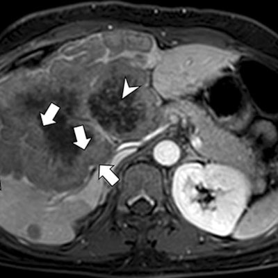
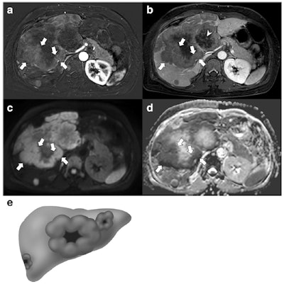 A 49-year-old woman with a gallbladder neuroendocrine tumor. A and B: On subtraction imaging of arterial phase and delayed phase of axial contrast-enhanced MRI, a 14.7-cm mass replacing the gallbladder and invading the liver is demonstrated. The mass shows thick rim enhancement with a thick peripheral enhancement and washout (arrows). Note the delayed central enhancement (arrowhead). On diffusion-weighted imaging (C) and apparent diffusion coefficient mapping (D), this mass also depicts a thick rim diffusion restriction (arrows). The diagnosis of neuroendocrine tumor was made via percutaneous biopsy. E: Thick rim appearance. All images courtesy of Drs. Jae Seok Bae, Se Hyung Kim, and colleagues and European Radiology.
"Neuroendocrine tumors and their metastases to the liver and lymph nodes more frequently demonstrated a thick rim appearance on contrast-enhanced MRI and diffusion-weighted images," they reported. "The ratio of apparent diffusion coefficient values between the lesion and the spleen was significantly lower for the primary mass, liver metastases, and lymph node metastases of GB-NETs than for those of GB-ADCs. A large metastatic lymph node was the only poor prognostic factor for overall survival in patients with GB-NETs and GB-ADCs."
Distinctive MRI features
Neuroendocrine tumors are rare and can develop in many different organs of the body. They affect the cells that release hormones into the bloodstream (neuroendocrine cells), and these tumors can be malignant or benign and they often grow slowly.
In the study, the authors' main objective was to identify MRI features that are helpful for differentiating GB-NETs from GB-ADCs and to evaluate their prognostic values.
Between January 2008 and December 2018, they retrospectively enrolled patients who underwent MRI for gallbladder malignancy. Two radiologists independently assessed the MRI findings and reached a consensus. They identified significant MRI features that distinguish GB-NETs from GB-ADCs.
Of 63 patients, 21 had GB-NETs and 42 had GB-ADCs. Compared with GB-ADCs, GB-NETs more frequently demonstrated well-defined margins, intact overlying mucosa, and thick rim contrast enhancement and/or diffusion restriction on MRI scans. Liver metastases were more common and demonstrated thick rim contrast enhancement and diffusion restriction in GB-NETs, while lymph node metastasis showed thick rim diffusion restriction more often in GB-NETs than in GB-ADCs, the authors reported.
On quantitative analysis, the sizes of the gallbladder mass and metastatic lymph nodes in GB-NETs were larger than those in GB-ADCs (p = 0.002 and p = 0.010, respectively). The ratio of apparent diffusion coefficient values between the lesion and the spleen was lower in the gallbladder mass, liver metastases, and lymph node metastases of GB-NETs than those of GB-ADCs (p < 0.001, p = 0.017, and p < 0.001, respectively).
Survival analysis revealed that a large metastatic lymph node (hazard ratio, 1.737; 95% confidence interval, 1.112-2.712) was the only poor prognostic factor (p = 0.015).
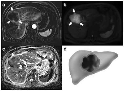 A 76-year-old man with gallbladder adenocarcinoma. A: Subtraction images taken during the arterial phase of an axial contrast-enhanced MRI show a 7.5-cm mass replacing the gallbladder with thin peripheral rim enhancement (arrows). On diffusion-weighted imaging (B) and apparent diffusion coefficient mapping (C), this mass demonstrates a nonthick rim appearance (arrows). Extended right lobectomy was performed, and a poorly differentiated adenocarcinoma was histopathologically confirmed. D: Nonthick rim appearance.
"We found that GB-NETs were significantly larger, frequently had a well-defined margin and intact overlying mucosa, and more often showed thick rim contrast enhancement and diffusion restriction, compared with GB-ADCs," the authors wrote. "In addition, liver and lymph node metastases were more common for GB-NETs, and these also demonstrated thick rim contrast enhancement and diffusion restriction."
In terms of clinical outcome, the study demonstrated that patients with GB-NETs had significantly shorter median overall survival than those with GB-ADCs: 12 months versus 44 months (p = 0.006).
A major limitation of the study was the small sample size, especially for GB-NETs, which was mainly due to their rarity, they explained. There was the potential for selection bias due to the retrospective nature of this study. Also, various MRI machines were used, including two different magnetic field strengths (1.5 and 3 tesla), which could have led to increased variability in apparent diffusion coefficient values; however, this reflects clinical practice and may increase the generalizability of the results.
"Radiologic-pathologic correlations could not be performed in many patients because surgery was contraindicated in those cases due to an advance stage of disease. Therefore, further studies are needed to investigate histopathologic backgrounds for our new findings such as intact overlying mucosa or thick rim appearance of GB-NETs," the team concluded.
