Myocarditis remains underdiagnosed, and lowering the threshold for diagnostic workup with cardiac MRI can help to identify myocarditis early, leading to significant improvements in patient care, according to a joint Swiss-German study published on 3 July in Swiss Medical Weekly.
By redesigning their approach to use cardiac MRI to examine all unexplained cases of myocardial infarction and nonobstructive coronary artery disease, and by implementing key MR imaging protocols during the scans, researchers were able to significantly increase the detection rate of myocarditis from 12.7 per 100,000 hospitalizations between 2011 and 2015 to 62.5 per 100,000 hospitalizations between 2016 and 2017.
"We have demonstrated that systematic workup of patients with angina-like symptoms, elevated troponin, and no significant coronary artery disease led to an almost fivefold increase in the diagnosis of myocarditis in our tertiary care center," wrote the researchers led by Dr. Bettina Heidecker of University Hospital Zurich. "These data suggest that myocarditis remains underdiagnosed and that lowering the threshold for diagnostic workup with cardiac MRI may help to identify myocarditis early."
Myocarditis is an inflammatory heart disease that can lead to dilated cardiomyopathy, arrhythmias, and sudden cardiac death. While cardiac MRI is one option to diagnose the condition, the "true incidence of myocarditis remains unknown," the researchers noted. "Postmortem studies have identified myocarditis in up to 42% of cases of sudden cardiac death, suggesting a significant epidemiological impact."
Naturally, earlier detection of any malady can lead to more effective treatment and greater chances of survival. "Timely and accurate diagnosis is crucial as certain subtypes of myocarditis require specific therapy," Heidecker and colleagues wrote.
Before changing their strategy, the researchers only used cardiac MRI in patients with classic symptoms of myocarditis, which include chest pain, dyspnea, and palpitations. Beginning in 2016, cardiac MRI was used for all unexplained cases of myocardial infarction and nonobstructive coronary artery disease, based on guidelines from the European Society of Cardiology (ESC).
They hypothesized that the rate of myocarditis detection would increase by 2017, "when cardiac MRI was systematically applied, compared to the detection rate between 2011 and 2015, when cardiac MRI was performed only when there had been moderate to high clinical suspicion of myocarditis."
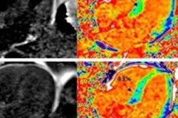
The retrospective, seven-year, single-center study included 556 patients (mean age, 57 years; range, 19-92 years) who underwent cardiac MRI on a 1.5- or 3-tesla scanner (Skyra, Siemens Healthineers or Achieva, Philips Healthcare) for diagnostic workup of angina-like symptoms and nonobstructive coronary artery disease (stenosis of less than 50%). The median time between symptom onset and scanning was 10 days (range, 4-23 days). The cardiac MR imaging protocol included T2-weighted imaging to assess edema and late gadolinium enhancement images acquired 10 minutes after weight-based administration of gadobutrol (Gadovist, Bayer HealthCare) to detect necrosis.
With the new approach, the researchers diagnosed 52 cases of myocarditis between 2016 and 2017, more than double the 24 cases of myocarditis between 2011 and 2015. The increase in the annual detection rate of 62.5 per 100,000 hospitalizations in 2016 and 2017, compared with 12.7 per 100,000 hospitalizations from 2011 to 2015, was a statistically significant difference of 4.9 in the detection ratio (p < 0.0001).
"The finding of a 4.9-fold increase in the total detection rate of myocarditis between 2016 and 2017 based on cardiac MRI and clinical findings compared to previous years suggests that myocarditis continues to be underdetected," Heidecker and colleagues concluded. "This supports the notion that diagnostic workup tools have to be improved."
