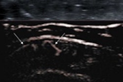
Contrast-enhanced ultrasound (CEUS) can be used instead of CT for following up solid-organ injuries in children and young adults, avoiding unnecessary radiation exposure in this vulnerable population, according to researchers from London's King's College Hospital who reported their findings from a large retrospective study.
The group found that CEUS detected all of the same complications that CT did on follow-up imaging. What's more, CEUS found several complications that were missed on CT.
"Our initial experience shows that CEUS diagnostic accuracy equals that of CT," said Dr. Annamaria Deganello. "We could correctly diagnose all complications that were seen on CT, but we could do it in a safe, radiation-free way [and that's] very important for these children."
She presented the research during a session at the recent American Institute of Ultrasound in Medicine (AIUM) meeting.
What's the best follow-up?
Conservative management is the most widely accepted strategy for most solid-organ injuries, and this approach requires appropriate follow-up imaging, Deganello said.
In the hope of reducing unnecessary radiation exposure in children and young adults, the researchers sought to compare the performance of CEUS with CT for following up such patients who have blunt or penetrating solid-organ injuries.
Deganello and colleagues retrospectively reviewed all referrals between 1999 and 2013 to the King's College Hospital level I trauma center, ending up with 766 consecutive patients who had received CT after sustaining abdominal trauma. There were 161 females and 605 males, with a mean age of 15 years (range, 9 months to 20 years).
Among the patients, 51% had a penetrating injury, 39% had been in a road traffic accident, 7% were injured in a fall, and 3% had other blunt trauma. The liver (35%), spleen (28%), and kidneys (17%) were the most commonly injured organs.
All patients received CT scans with a postcontrast split-bolus (for those who had blunt abdominal trauma) or dual-phase protocol (for those who had penetrating injuries). Contrast ultrasound scans were performed by experienced radiologists after obtaining informed consent from adult patients and parents, Deganello said.
King's College Hospital uses a split-screen approach to CEUS for follow-up of trauma patients: B-mode images are displayed on one half of the screen while CEUS images are shown on the other half, she said. A bolus of SonoVue (Bracco Diagnostics) is injected through peripheral intravenous access with a dose of 0.5 mL to 2.4 mL, depending on the size of the child. Both cine loops and static images are stored.
Of the 766 patients, 112 had at least one follow-up CT scan for solid-organ abdominal injury, and 37 (33%) also had a follow-up CEUS scan. In all of those patients, CEUS findings matched those of CT, and CEUS correctly diagnosed all complications when compared with CT, Deganello said.
In addition, in three of the 37 cases, CEUS diagnosed lesions that were not seen on CT, including a splenic pseudoaneurysm, a grade 1 renal contusion, and a grade 2 splenic contusion.
"In those cases where CT was still performed despite [CEUS showing] reassuring resolving injuries, CT never showed any new injuries compared with CEUS," she said.
In 2011, contrast ultrasound was introduced into the institution's follow-up pathway for solid-organ injuries. Since then, 30 (40%) of 75 patients have been followed up only with CEUS, Deganello said. All of these patients fully recovered without further complications, including one with a pseudoaneurysm that was managed conservatively.
Although the use of contrast ultrasound in children is considered to be "off-label," there have been no adverse reactions, she noted.
"CEUS can really be a viable alternative to CT for the follow-up of [solid-organ] injuries in this vulnerable population," she concluded.


