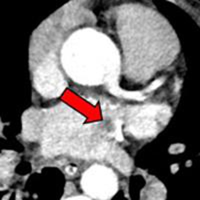
Echocardiography may be the de facto gold standard for cardiac evaluation of patients suspected of having cardioembolic stroke, but it isn't necessarily the best modality. A new study from Saudi Arabia used CT to discover several thrombi that echo had missed, along with other conditions.
While the study of more than 350 patients with suspected cardioembolic stroke didn't calculate sensitivity or specificity, it did show dozens of potentially significant findings at CT that were not visualized at echocardiography, said Dr. Amr Ajlan and his colleague Dr. Rabab Bagdadi, radiologists from the cardiothoracic imaging unit at King Abdulaziz University Hospital in Jeddah.
"Cardiac CT adds value to echocardiography in detecting major embolic potential causes, and it also might add some value in detecting stroke-unrelated findings and coronary atherosclerosis," Ajlan said in a presentation at ECR 2015.
 Above, an adult with a left atrial thrombus that was unreported on echocardiography and seen on CT as a filling defect on the arterial (red arrow) and delayed (orange arrow) enhancement phases. Below, an adult with remote inferior myocardial infarction that was unreported on echocardiography and seen on CT as left ventricular inferior myocardial thinning (red arrow). All images courtesy of Dr. Amr Ajlan.
Scanning for cardioembolic stroke
King Abdulaziz University Hospital started providing neuroradiology services of cardiac CT for cardioembolic stroke about 2.5 years ago, Ajlan said, and the new study was designed to see the effects of this practice. Ajlan and Bagdadi retrieved all cardioembolic stroke cases from the hospital's PACS from June 2012 to December 2014; this consisted of 357 cardiac cases, of which 47 were neurology referrals.
Using a dual-source, 128-detector-row scanner (Somatom Flash, Siemens Healthcare) and a same-day protocol, the patients underwent CT after echocardiography for suspected cardioembolic stroke. Some 72% of the CT scans were acquired retrospectively, with the rest done prospectively, and 86% of cases included the aortic arch.
 Dr. Amr Ajlan.
One of three experienced cardiothoracic radiologists read the images retrospectively and correlated the findings with echocardiography reports, attempting to provide a potential explanation when cardiac CT showed an important finding.
Per European Society of Cardiology guidelines, readers focused on findings of major embolic potential, including thrombi, tumors, vegetations, aortic atheromas, evidence of myocardial infarction, left ventricular aneurysm, mitral valve disease, shunts, and vasculitis, Ajlan said. They also looked for significant findings unrelated to stroke, such as atherosclerosis.
The 357 patients (mean age, 52 ± 11 years) had high rates of diabetes (30.8%) and hypertension (72.3%), both of which are risk factors for cardioembolic stroke.
Finding what echo missed
CT found five thrombi missed by echocardiography; they were located in the left atrial body, the left atrial appendage, the left ventricular apex, a repaired mitral valve, and the aortic arch. CT did miss a thrombus at the tip of the left atrial appendage that was visualized on echocardiography and seen only retrospectively on CT.
"I think we think missed this because we did not include delayed imaging in this case," Ajlan said. "The reader probably thought it was within an adjacent cardiac recess."
CT discoveries unrelated to stroke included three pulmonary emboli, two cases of aspiration pneumonia, two cavitary pneumonias, one septic embolus, one lymphadenopathy, one lymphatic mass, one ankylosing spondylitis, one case of hiatal hernia and esophageal thickening, and three other findings.
Consistent with the high prevalence of diabetes and hypertension in the patient population, 23 (49%) of the 47 patients had unexpected coronary artery disease at CT, and significant coronary artery stenoses (≥ 50% luminal narrowing) were found in 54% of patients.
 Above, an adult with indeterminate vasculitis that was unreported on echocardiography and seen on CT as mural aortic thickening (orange arrow) with extension into arch vessels (red arrow). Below, an adult with a left atrial appendage tip thrombus that was missed on CT and subsequently detected on echocardiography. The abnormality was noted on CT in retrospect (red arrow).
Study limitations included its retrospective and single-center design. In addition, the researchers relied on echocardiography reports, and most of the echo was transthoracic echocardiography, Ajlan said.
"What can we learn from this? This is a descriptive study, not a study of diagnostic accuracy," he said. "No. 2 is that CT is a very appealing modality."
CT as emerging standard of care?
Transesophageal echocardiography usually served as the gold standard in the hospital; "however, it's logistically very difficult," he continued. "You need anesthesia, you need protection; you need time and expertise to do that."
At the beginning of the study, most patients underwent transesophageal echo, but by the end, only some had transesophageal echo, and in one or two cases echo was skipped entirely.
"So the neurologists were changing their practices according to what they saw on the CT images," Ajlan said.
And while it's too early to say that cardiac CT is now the preferred test, it does add value for detecting potential causes of cardioembolic stroke, as well as in revealing findings unrelated to stroke.


