Detailed high-resolution 3D 256-detector-row CT images have opened a fascinating window onto the life of a teenager who lived in what is now northern Israel and who suffered severe head trauma 100,000 years ago, according to a French anthropologist.
The injuries seen in the adolescent Paleolithic skull from the lower Galilee region are characterized by an anterior depression on the right side of the frontal squama and are likely to have had a severe impact on the unfortunate youth's speech and social functioning. But signs of an unusual ritual burial suggest the special esteem in which the 12- or 13-year-old was held by those in the community.
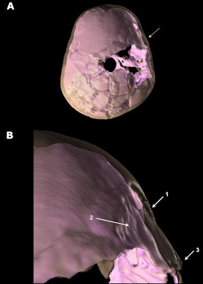 Above, superior view of Qafzeh 11 3D reconstructed skull showing the depressed fracture on the frontal right side. The skull vault appears in transparency and the virtual endocranial cast in pink. A: General view. B: Close-up view of the trauma area. 1: anterior part of the frontal bone depressed fracture penetrating the endocranial volume. 2: irregular shape of virtual endocranial surface indicating brain damage. 3: diastasis of the right coronal suture. Images republished courtesy of PLOS One.
Whether the youth was attacked or injured accidentally can't be determined, despite the 3D CT images of the skull, noted lead author Hélène Coqueugniot, PhD, research director at the French National Center for Scientific Research in Pessac, France. Coqueugniot is also an anthropologist at the University of Bordeaux and at the Max Planck Institute for Evolutionary Anthropology in Leipzig, Germany.
The CT images tell us much about the youth's injuries.
"Thanks to 3D reconstructions that allowed us to visualize both the cranial vault and the endocranial cast, we were able to conclude that the frontal fracture was a severe one (depressed); that brain was damaged in the area of the fracture; and that probable secondary cognitive and behavioral troubles did occur," she explained in an email to AuntMinnieEurope.com.
Middle Paleolithic period remains
The Qafzeh site in the lower Galilee region has yielded the largest Levantine hominin collection from Middle Palaeolithic layers to be unearthed. The samples have been dated circa 90,000 to 100,000 years old or to marine isotope stage 5b-c, the authors noted. Within this collection, remains of the youth known as Qafzeh 11 include a skull lesion that was previously attributed to a healed trauma.
"Three-dimensional imaging methods allowed us to better explore this lesion, which appeared as being a frontal bone depressed fracture, associated with brain damage," Coqueugniot and colleagues wrote.
Interestingly, the endocranial volume of the skull was smaller than expected for the dental age, a finding that supports the hypothesis that growth may have been delayed due to traumatic brain injury (TBI). Even so, the trauma didn't affect the typical human brain morphology pattern of the right frontal and left occipital petalia, they noted (PLOS One, 23 July 2014).
"I wonder if the injury stunted 11's growth, or he/she was just small," Coqueugniot wrote in her email.
Troubled youth
Judging from the injuries seen on the CT scans, the youth quite likely suffered from personality and neurological problems directly related to the brain damage. Adding to the mystery, the adolescent was given a ritualized funeral that was unique among the southwestern Asian burials dated to the Middle Palaeolithic period.
There are two broad categories of changes that typically occur after brain trauma, Coqueungniot explained. First, there are the general troubles characterized by personality changes whenever there is focal injury, and this can manifest in distress or impairment in social or occupational areas of functioning.
Second, are the neurological disorders due to focal damage, in the areas of the brain responsible for psychomotricity, managing uncertainty, visual attention, and oral communication, she wrote.
"It is clear that this young individual suffered from marked troubles in social behavior," she wrote. "This individual was probably perfectly able to move, to walk, to eat, and drink alone -- but more complex tasks such as production of lithic artifacts, hunting strategy, tools, and weapons use, could have been difficult to perform."
Imaging the skull
CT scans of the Qafzeh 11 skull were performed to reassess the traumatic condition that affected the adolescent. The 3D reconstructions enabled precise visualization of the internal and external surfaces of the cranial vault, as well as inner structures in the area of the injury. They open a window into the potential impact of surface injury to the brain.
The scan was performed at Carmel Medical Center in Haifa, Israel using a Brilliance iCT 256 scanner (Philips Healthcare) with a voxel size of 0.67 mm. Acquisition parameters included 120 kV and 298 mAs.
The study team created 3D reconstructions of the skull and made an endocast from the scans. The 3D reconstructions were performed using a free software package, Treatment and Increased Vision for Medical Imaging (TVIMI), developed by the University of Bordeaux and based on a half maximum height (HMH) algorithm.
TVIMI software "proved to be more reliable for 3D measurements than other software programs currently implementing different algorithms for 3D reconstructions," the study team wrote. The software provided estimates of endocranial volume, and created virtual reconstructions that allowed the scientific team to localize the impact of the cranial lesion on the brain surface using the 3D reconstruction of the extant human brain and verifying the correspondence of the anatomical landmarks.
The problem with traditional commercial 3D software programs that provide "nice reconstructions" is that they can impart a smoothing effect that hides important details, Coqueugniot wrote in her email.
More severe than thought
The reconstructions showed clear evidence of a skull fracture of the right part of the frontal bone -- located on the right part of the frontal squama just above the petrionic region that was in the process of healing. The fractured quadrilangular fragment (about 30 x 23 x 26 x 12 mm) is near the frontal boss and the upper face at 40 mm from the frontal midline.
"This type of fracture that can be related to a blunt force trauma, clinically corresponds to cranio-encephalic wound and raises the question of its impact on the brain," the authors wrote.
Advanced CT changed the characterization of the youth's condition from mild disability to something that appears much more serious. Such an injury presents a high-risk level for brain damage, including intracranial hemorrhages, as well as nervous system lesions such as concussion, contusions, and lacerations, which can lead to brain tissue destruction or cerebral scar.
Lesions in the right frontal area (areas responsible for psychomotricity) may have led to problems controlling movement, performing specific tasks, managing uncertainty, and visual attention and eye movement, they continued. Possible injury to another area may have caused problems in oral communication.
"It is highly probable this young individual suffered also from personality changes due to traumatic brain injury," they wrote. Such personality disturbances occur in about 65% of individuals with similar mild to moderate injuries.
Ritual burial
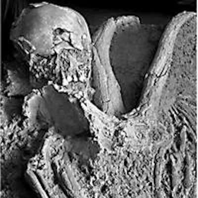
The adolescent was buried with two deer antlers placed on the upper part of the chest, near the palmar side of the hand bones. The particular hand location, in the context of the body spatial arrangement, was evidence of a funerary offering rather than an accidental inclusion.
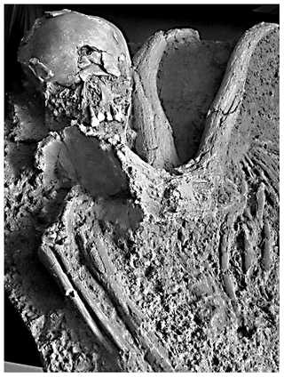 Above, partial view of the Qafzeh 11 burial showing the deposit of the red deer antlers in close contact with the child skeleton (cast).
Several other burials at the site were not so arranged, meaning that Qafzeh 11 represents a case of special treatment and convincing evidence for ritual behavior. This evidence demonstrates elaborate social behavior among the Qafzeh middle Paleolithic people.
"Actually, it is very difficult to reconstruct the social behavior of these ancient people who lived 100,000 years ago," Coqueugniot told AuntMinnieEurope.com. "We just have some landmarks such as, in this case, burial practice, which is special for an individual who was, during their life, somehow special after having experienced a severe TBI."
As a result, the researchers' hypothesize that the youth, "having both post-traumatic cognitive and social communication troubles was recognized by members of their group as being different," Coqueugniot noted. "They marked their recognition by burying this special individual with special care."
The team plans to continue its work on this specimen and others from the same site, eventually aiming to re-examine other middle Palaeolithic specimens from the region using 3D CT to reveal new information.


