While contrast-enhanced spectral mammography (CESM) is a relatively new technique, two Egyptian studies are showing the advantage of using the technology for the detection of breast lesions, and compare it favorably with more established imaging modalities -- in particular for imaging facilities with limited resources.
In research presented at ECR 2014, one study compared CESM with dynamic contrast-enhanced MRI for the detection and staging of breast cancer, finding it to be a comparative technique. A second study found the technology to be useful in detecting lesions in the edematous breast, a condition that can confound conventional mammography.
In addition to its comparable diagnostic performance, CESM could offer benefits over breast MRI based on cost, availability, and patient convenience, the authors said.
Better together
Study author and presenter Dr. Rasha M. Kamal, professor of radiology at Cairo University, told AuntMinnieEurope.com that in the field of breast imaging the best results are achieved from modalities that can combine both morphology assessment and functional information.
 Dr. Rasha M. Kamal (right) with Dr. Dorria Saleh El Sayed Salem (left), both of Cairo University in Egypt. El Sayed Salem was not involved in the current study. Image courtesy of Dr. Rasha M. Kamal.
While mammography is considered the gold standard in the field of breast imaging, its accuracy is limited, especially in dense breasts, Kamal explained. The addition of functional information can improve diagnostic accuracy, particularly for challenging cases.
Functional detail is usually obtained by injection of a contrast medium, she said, further noting that the only imaging modality that combined both was dynamic contrast-enhanced MRI (DCE-MRI). This is, until CESM was introduced.
Even when tumors are detected, the full extent of disease may not be clearly depicted. Growth of tumor cells requires the formation of new blood vessels (angiogenesis) to supply the oxygen and nutrients necessary for growth and survival. Therefore, imaging modalities coupled with contrast can potentially aid in the detection, diagnosis, and staging of breast cancer, she added.
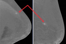
CESM involves the acquisition of two sets of images at different energy levels after the injection of contrast. The image at one energy level typically displays a conventional mammographic image, while the other energy level displays vascular information from angiogenesis that can be a sign of cancer.
CESM has evolved as an important adjunctive tool to mammography, is expected to increase mammography's accuracy, and can be used in the clarification of mammography-identified equivocal lesions.
"It can help in the assessment of the extent of disease for presurgical planning and has the potential to be used in monitoring of neoadjuvant therapy and assessment for tumor recurrence," Kamal said.
In a prospective study, Kamal and colleagues included 70 patients with 82 breast lesions, of which 47% (33) were benign and 53% (37) were malignant. The research was conducted at the National Cancer Institute -- Cairo University.
Both CESM and breast MRI were compared with regard to their ability in detecting and diagnosing breast cancer, and when cancer was diagnosed they were compared in their ability to stage. Specifically, the researchers looked at how far the cancer had spread by determining size of breast lesions, multicentricity, and multifocality, as well as bilaterality. Also, they looked at lymph node status and distant metastases.
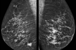
Standard digital mammography was performed in the mediolateral oblique and craniocaudal projections, followed by CESM at low (22 kVp to 33 kVp) and high (44 kVp to 49 kVp) energy exposures in the same projections. MRI was set in the axial orientation and postprocessed using maximum intensity projection (MIP) and multiplanar reformat (MPR) images.
The detection and diagnosis of disease were independent on breast density in both modalities. While both showed accurate lesion size identification as compared with lesion sizes in pathology specimens, contrast mammography was highly accurate in the identification of multifocal, multicentric, and bilateral disease, which is an essential step before therapy planning, Kamal noted. Meanwhile, a major advantage of MRI was its ability to identify anterior chest wall invasion.
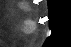
CESM was superior to MRI in the identification of tiny malignant satellite tumor foci. In the contest of malignancy, both modalities stood on the same level. Enhancement detection of some benign lesions was limited in contrast mammography.
Statistical analysis yielded a sensitivity, specificity, and accuracy of 93.7%, 66.6%, and 80.6% for contrast mammography, compared with 93.7%, 85%, and 90.3% for MRI. So while both modalities showed comparable sensitivity, MRI showed a higher specificity because of the large number of false-positive cases on contrast mammography.
"One of the main reasons for false-positive results was a ring pattern of contrast uptake, which includes a wide spectrum of pathologies, from benign cases of mastitis with abscess formation to frank malignant lesions," Kamal explained.
And while both modalities had comparable indications, contrast mammography had fewer contraindications. For example, while renal impairment was a concern with both, pregnancy was only a concern with contrast mammography, while comfort, weight, and claustrophobia were all concerns linked to MRI.
Kamal noted that contrast mammography is a 2D imaging modality that is still subject to some compromise due to tissue overlap, in comparison to MRI, which allows clinicians to assess the morphological criteria of breast lesions in different pulse sequences together with kinematic assessment.
Contrast mammography's advantages
She explained there are several advantages to using contrast mammography, because not only is it economical but also presents a major advantage in terms of time saving due to a long waiting list on MRI schedules -- because MRI is not just dedicated to breast examinations. Also, contrast mammography can be performed on the same day as the initial mammography study, which reduces patient anxiety.
Contrast mammography is also very easy to perform, and much cheaper, which is a point to be highly considered in the presence of limited resources, Kamal said.
Kamal noted she has been working on contrast mammography for two years now and recently introduced a contrast mammography machine in Egypt's national breast cancer screening program, in which she is the assistant project manager.
"We are also in the process of fixing another machine in Cairo University Hospital in the near future," she added.
Second study
Another study at ECR 2014, presented by Dr. Noha ElSaid, an assistant professor of radiology at Cairo University, found that contrast mammography is useful in detecting lesions in the edematous breast. She also noted that the technique is a simple way to enhance the detection and characterization of breast lesions, especially in dense breasts.
ElSaid and colleagues looked at 36 patients (age ranged from 37 to 62 years) who presented with swollen breasts. The study subjects underwent conventional mammography, contrast mammography, and ultrasound breast examinations.
The researchers found the sensitivity and specificity of contrast mammography to be 92% and 86.7% versus 88% and 76.5% for conventional mammography.
"Dual-energy contrast-enhanced digital mammography is a useful technique in identification of lesions in mammographically dense edematous breasts and is capable of demonstrating lesions that are not visible at standard mammography," the authors wrote.
In conclusion, they noted that contrast mammography is useful in follow-up of cases presenting by edema after conservative breast surgery and chemotherapy.


