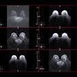
Low-dose stress CT myocardial perfusion imaging (CTP) of the heart is feasible at a tube current of 80 kV, producing image quality equivalent to 100 kV in a group of nonobese patients with a lower radiation dose, reported a team from Japan in European Radiology.
Specifically, stress CTP imaging set at 80 kV and 370 mAs cut radiation dose by 40% compared with the same exam at 100 kV and 300 mAs, according to radiologists from Japan. Even so, signal-to-noise ratio (SNR), contrast-to-noise ratio (CNR), and myocardial blood flow (MBF) were comparable with the standard-dose exam, wrote the study team.
The low-dose exam was "associated with a nearly 40% reduction in radiation dose, no significant difference in SNR and CNR, and comparable estimated MBF value compared with CTP using 100 kV," wrote Dr. Makiko Fujita and colleagues from Mie University School of Medicine in Mie, Japan (Eur Radiol., March 2014, Vol. 24:3, pp. 748-755).
The price to pay for CT
Stress CT myocardial perfusion is an increasingly popular choice to access myocardial function, as it goes beyond coronary CT angiography in its ability to assess the functional effects of stenosis. But there's a price to pay for the additional information.
"Dynamic myocardial perfusion imaging results in a relatively high absorbed radiation dose to the patients," the study team wrote, noting a mean radiation dose for 128-detector dual-source CT MPI ranging from 9.2 mSv to 12.5 mSv using tube voltage of 100 kV and tube current of 300 to 330 mAs.
The study sought to determine the image effects of reducing the settings from 100 kV and 370 mAs to 80 kV and 300 mAs, as well as any differences in the myocardial blood flow calculated from CTP images in patients with BMI of 25 kg/m2 or under.
The researchers examined 30 consecutive patients ranged from 45 to 85 years in age (mean age 67 years, mean BMI 21 kg/m2) who were referred for dynamic myocardial CTP with adenosine stress during the first nine months of 2012. The patients were clinically referred for coronary CT angiography (CTA) for known or suspected coronary artery disease (CAD). Excluded were patients after hypertropic cardiomyopathy, after coronary artery bypass graft (CABG) procedures, and those with implanted pacemakers, impaired renal function, or contraindications to iodinated contrast.
Following administration of 370 mgI/mL iopamidol (Iopamiron 370, Bayer Healthcare Pharmaceuticals) at 5 mL/sec, patients were scanned using dual-source CT (Somatom Definition Flash, Siemens Healthcare). The 30 patients were randomized (15 each) to either 80 kV and 370 mAs or 100 kV and 300 mAs. Dynamic stress images were acquired in 1-mm increments over 30 seconds to visualize the myocardium, followed by prospectively gated coronary CT angiography (CCTA) in 0.75-mm increments to visualize the coronary arteries, the study team wrote.
The investigators started the dynamic myocardial CTP scan six seconds before arrival of the contrast medium bolus in the ascending aorta. They initiated myocardial CTP three minutes after administration of adenosine at 0.14/mg/kg/min. Dynamic data were acquired using ECG-triggered axial acquisitions repeated at two table positions for z-axis coverage of 73 mm, the group wrote.
Fujita and colleagues reconstructed images every 1 mm using a B23 kernel, which includes beam-hardening correction, and examined them on a perfusion imaging workstation (Syngo VPCT body, Siemens). MBF was estimated using a parametric deconvolution technique, and the algorithm generated an MBF map.
According to the results, scanning with the 80-kV technique versus 100-kV reduced radiation dose by 40% (mean dose-length product [DLP] 359 ± 66 for 80 kV versus 628 ± 112 mGy cm for 100 kV, p < 0.001). At the same time, both CNR (34.5 ± 13.4 for low-dose versus 33.5 ± 10.4 for 100 kV, p = 0.81) and MBF in the nonischemic myocardium (0.95 ± 0.20 for 80 kV versus 0.99 ± 0.25 ml/min/g of 100 kV; p = 0.66) remained essentially unchanged, the group reported.
Scans acquired at 80 kV had maximal enhancement that was 30.9% higher than 100 kV (804 ± 204 HU for 80 kV versus 614 ± 115 HU for 100 kV; p < 0.005). However, the reduced-dose scans also had 31.2% greater noise (22.7 ± 3.5 for 80 kV versus 17.4 ± 2.6 for 100 kV; p < 0.001).
| Results summary in 30 patients | |||
| 80 kV (n = 15) | 100 kV (n = 15) | P value | |
| DLP (mGy cm) | 359 ± 66 | 628 ± 12 | < 0.001 |
| Estimated effective dose (mSv) | 6.1 ± 1.1 | 10.7 ± 1.9 | < 0.001 |
| Size-specific dose estimate (mGy) | 71.1 ± 14.5 | 123.7 ± 26.4 | < 0.001 |
| Image noise (HU) | 22.7 ± 3.5 | 17.4 ± 2.6 | < 0.001 |
| SNR | 2.4 ± 0.7 | 2.9 ± 0.8 | 0.09 |
| CNR | 34.5 ± 13.4 | 33.5 ± 10.4 | 0.81 |
| Mean MBF (mL/min/g) | 0.95 ± 0.20 | 0.99 ± 0.25 | 0.66 |
Dynamic CT has distinct advantages over static CT, in that it "allows for noninvasive quantification of MBF from time-attenuation curves of both the blood pool and the myocardium," Fujita and colleagues wrote. "However, dynamic CTP is associated with a higher absorbed radiation dose to the patients in comparison with static CTP."
The authors said they were aware of only one previous study of dose reduction in dynamic myocardial CTP. That study compared two dynamic CTP protocols with and without automated tube current modulation, but the tube voltage was fixed at 100 kV for both arms. The use of tube current modulation reduced dose by 36% for a mean effective radiation dose of 7.7 ± 2.5 mSv for the lower-dose exam -- and again without deterioration of SNR and CNR.
The authors pointed out the study limitation of requiring all patients to have a BMI of 25 or less, and noted that a new integrated circuit detector may enable scanning of overweight patients at 80 kV in a future study. They also stated that MBF values were not validated against a gold standard such as radio-water PET in the present study, but that the results of an animal experiment suggest that the derived MBF figures they used were accurate.
"The use of 80-kV tube voltage should be considered in dose-reduction strategies for dynamic CTP of individuals with a normal BMI," Fujita and colleagues wrote.
