PET/MRI is more than adequate in characterizing liver lesions and provides greater lesion conspicuity than PET/CT, offering clinicians a powerful alternative for oncology imaging, according to a German study published in the November issue of the European Journal of Radiology.
PET/MRI provided better diagnostic confidence due to soft-tissue contrast and complementary information from different MRI sequences, according to lead author Dr. Karsten Beiderwellen and colleagues from University Hospital Essen, University of Duisburg-Essen, and University of Dusseldorf (EJR, Vol. 82:11, pp. e669-e675).
"The inherent soft-tissue contrast coupled with the higher spatial resolution of MRI is the main advantage over PET/CT, providing the overall higher conspicuity of liver lesions," Beiderwellen wrote in an email to AuntMinnie.com. "Plus, diffusion-weighted [MRI] helps in delineating even small lesions and offers complementary information to PET."
The study is the first to investigate the efficacy of simultaneous PET/MRI acquisition for liver lesions and metastases, he added.
More accurate differentiation
For many years, CT and transabdominal ultrasound have been used regularly in tumor staging, but the introduction of whole-body FDG-PET/CT has provided more accurate differentiation of lesions due to the glucose metabolism information provided, according to the authors.
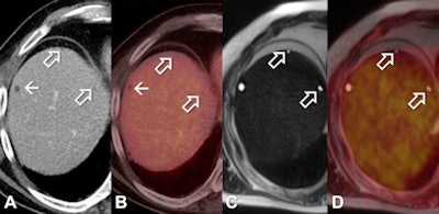
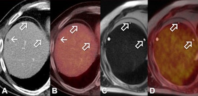 Patient with liver cyst in the liver segment VII identifiable both in PET/CT (A, B) as well as in PET/MRI (C, D). Additional small hypodense liver lesions in segment VIII deemed too small to characterize in PET/CT. In the T2-weighted half-Fourier acquisition single-shot turbo spin-echo (HASTE) in PET/MRI (C, PET/MRI fusion) the lesions exhibit a high signal and well-defined lesion borders and can therefore correctly be identified as liver cysts. Images courtesy of Dr. Karsten Beiderwellen.
However, liver lesions with a diameter of less than 15 mm are often considered too small to characterize on CT and are present in as many as one in three patients with liver lesions. The differentiation of small liver lesions is also a concern due to the limited spatial resolution of PET.
By comparison, MRI offers enhanced soft-tissue contrast and has proved particularly beneficial for patients with small liver lesions. The modality has shown superior results compared to CT and PET/CT for depicting and characterizing liver tumors, Beiderwellen and colleagues wrote.
"While PET/CT already represents a reference method in whole-body staging of cancer patients, combined PET/MRI might advance to become a possible future alternative," they wrote.
For the current study, the researchers prospectively enrolled 70 patients (31 women and 39 men with a mean age of 56 years ± 14 years). The subjects had confirmed solid tumors, were scheduled for whole-body FDG-PET/CT, and gave consent for an additional whole-body PET/MRI study.
Subjects were excluded if they were younger than 18 and had contraindications to MRI, such as an implanted device or claustrophobia. Primary tumors included 25 malignant melanomas, 10 breast cancers, and nine lung cancers.
Whole-body PET/CT was performed on a 128-slice CT scanner (Biograph mCT, Siemens Healthcare) with an axial field-of-view of 21.8 cm. PET imaging started 60 minutes after FDG injection of 291 ± 46 MBq and included five to six bed positions at approximately two-minute intervals.
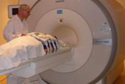
Participants then underwent simultaneous PET/MRI on a 3-tesla system (Biograph nMR, Siemens) with an axial field-of-view of 25.8 cm. The PET/MRI scan started 155 minutes (± 51 minutes) after FDG injection and included a whole-body PET scan with five to six bed positions at eight minutes each from skull base to midthigh with 3D image reconstruction.
Two experienced readers independently reviewed the images and rated lesions for PET/CT and PET/MRI as well as for each submodality. Malignant disease was confirmed by fine-needle aspiration or surgical intervention, but histopathological correlation for every detected liver lesion was not available.
Lesion detection
Liver lesions were present in 36 (51%) of the 70 patients based on the reference standard: 10 patients (14%) had liver metastases and 26 (37%) had benign liver lesions.
Both PET/CT and PET/MRI correctly identified all 10 patients with liver metastases. CT and MRI alone discovered all 10 patients with liver metastases, whereas PET alone identified nine patients.
A total of 97 lesions were found among the study cohort, with 26 lesions characterized as malignant and 71 lesions as benign. PET/MRI depicted all 97 liver lesions correctly, but PET/CT missed nine benign lesions.
However, additional malignant liver lesions were not found using PET/MRI, the authors noted; both PET/CT and PET/MRI detected the 26 malignant lesions. CT alone identified 87 total lesions (25 malignant and 62 benign), while MRI correctly identified all 97 benign and malignant lesions.
PET performed better when matched with MRI, according to Beiderwellen and colleagues. PET alone from PET/CT detected 14 (54%) malignant lesions, compared with 21 (81%) malignant lesions from the PET component of PET/MRI.
Key findings
The authors pointed out two key conclusions from the study. First, because integrated whole-body PET/MRI offers higher lesion conspicuity than PET/CT, more lesions can be delineated in PET/MRI.
"Second, PET/MRI offers higher diagnostic confidence in the characterization of liver lesions," they wrote. "Due to the excellent soft-tissue contrast and complementary information from different MRI sequences a correct differentiation especially of small liver lesions is possible."
An additional clinical benefit for patients is less radiation exposure with PET/MRI compared to PET/CT, Beiderwellen said.
The authors did note study limitations, including the small number of patients with liver metastases. Thus, the results "may only be considered as proof of potential of the method," they wrote.
"Despite its limitations, our study positively demonstrates the high potential of PET/MRI for the depiction and characterization of liver lesions" they concluded. "When combined with high-resolution sequences of the liver, PET/MRI with F-18 FDG might advance to a powerful alternative in oncologic imaging."
The group plans to expand the research to larger, prospective studies to evaluate the full potential of PET/MRI for liver imaging.
"Liver metastases in PET/MRI are a hot topic," Beiderwellen told AuntMinnie.com, "so we will have a further look in larger-scale studies on accuracy as well as clinical impact. Furthermore, we are evaluating the performance in malignancies of the female pelvis, where the method delivers very promising results."

