Researchers from the University of Aberdeen in Scotland have received a grant of 979,000 pounds (1.16 million euros) to develop a new MRI technique known as zero-field MRI, which could detect disease at earlier stages than current MR techniques.
The project is a collaboration of University of Aberdeen researchers in medical physics, radiology, neuroscience, and neurology, who are building the technology on-site. The project isn't the first groundbreaking MRI development; Aberdeen scientists and physicians were the first to scan a patient's body using MRI in 1980, the university said.
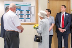
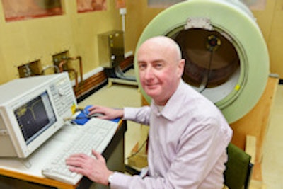
 Ready for action: David Lurie, PhD, professor in biomedical physics at the University of Aberdeen.
Chief investigator and medical physicist David Lurie, PhD, said neurodegenerative diseases are a serious concern, with more than 850,000 cases in the U.K., and numbers are expected to double in the next 40 years due to the aging population. He said the need for new drugs to slow disease progression is urgent, but first, imaging methods are needed to detect early brain changes before the onset of symptoms.
Technologies such as zero-field MRI could potentially fill the bill by enabling earlier disease detection. Unlike conventional MRI scanners, zero-field MRI would lower the magnetic field within the scanner down to almost 0 in order to see disease-related tissue changes that cannot be seen in conventional MRI, he said.
Along with neurodegenerative disease, zero-field MRI has great potential for diagnosing other diseases such as cancer, osteoarthritis, fibrosis, and thrombosis. The current project will take three years and focus on modifying the institution's fast-field cycling MRI scanner to allow zero-field measurements, the researchers said.
The research team will begin by scanning small objects such as bottles containing protein gels to mimic normal and diseased tissues. This work will be followed, hopefully, by the imaging of some patients with neurodegenerative diseases, the study team wrote.
