The ability of diffusion-weighted imaging (DWI) to create good reproducibility, correlate with histopathology, and enhance sensitivity to assess treatment results may make MRI an "excellent tool" to monitor response in metastatic colorectal cancer patients, according to a new Dutch study.
As apparent diffusion coefficient (ADC) values become increasingly important oncological biomarkers, researchers from Radboud University Nijmegen Medical Centre in the Netherlands found that DWI provides "excellent reproducibility" in ADC values compared to other imaging modalities. The clinical benefit is a more efficient method to monitor the progression of colorectal cancer treatment.
Lead study author is Dr. Linda Heijmen from the university's department of medical oncology and her article was published online on 23 September in the European Journal of Radiology.
Colorectal cancer is the second leading cause of cancer-related death in Europe, according to an April 2012 study in the Annals of Oncology, she noted. As in most cases of cancer, early detection can improve response to treatment and patient survival.
Previous research has shown that DWI-MRI can help characterize tissue, predict a response to treatment, and evaluate cancer. DWI measures ADC values, which relate to the movement of water molecules in vivo and indirectly reflect tissue microstructural characteristics.
Patient cohort
The Dutch study recruited 19 patients with liver metastases of colorectal cancer who were scheduled for metastasectomy between August 2009 and January 2011. The research excluded patients with a contraindication to MRI, such as claustrophobia, incompatible implants, and pacemakers. Patients received chemotherapy until one month before they joined the study. No chemotherapy or other therapy was administered between the two DWI examinations.
Respiratory-triggered DWI was performed twice within one week on patients scheduled for metastasectomy of colorectal liver metastases. All patients underwent surgery within three weeks of their last lesion measurement. The measurements were conducted on a 1.5-tesla whole-body MRI (Magnetom Avanto, Siemens Healthcare) with a body coil for excitation and a six-channel body matrix coil combined with a 24-channel spine matrix coil for signal reception.
Researchers drew a region-of-interest on all slices with a visible tumor. In cases where patients received an FDG-PET scan, researchers used those images to definitively determine if a lesion were a cyst rather than a liver metastasis. The study cohort included 23 metastases, which were detected through DWI in 19 patients. The average size of the metastases was 31.3 cm3 (range 0.7 to 130.4 cm3).
DWI located 18 metastases in the right liver lobe and five metastases in the left liver lobe. Three patients who completed both MRI scans did not undergo the planned metastasectomy, one metastasectomy was changed to radiofrequency ablation, and two patients had unresectable metastases, making histological data available for 16 patients.
DWI reproducibility
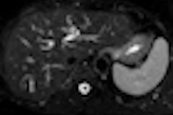
In their DWI analyses, Heijmen and colleagues found that liver metastases were commonly visible on ADC maps as hypointense lesions. The mean ADC value in all liver metastases was 1.17 (± 0.11) × 10-3mm2/s. The coefficient of reproducibility for the mean ADC value of the tumor, based on a lesion-by-lesion analysis of 21 metastases that were imaged twice, was 0.20 × 10⁻³mm2/s.
In addition, the authors noted the "reproducibility of the ADC mean did not depend on the volume of the metastases, previous treatment, or the location in the right or left liver lobe."
The mean ADC value of the lesions in patients pretreated with systemic therapy also was found to be greater when compared with the ADC of untreated lesions. The higher ADC values, the study stated, were more prominent in the most recently treated patients. Metastases in patients who had received their last systemic treatment within three months of the first DWI had a mean ADC of 1.27 ×10⁻³mm2/s, compared with 1.05 × 10⁻³mm2/s in previously untreated tumors.
Hence, Heijmen and colleagues concluded DWI in liver metastases "showed excellent reproducibility, both compared with other imaging techniques and compared with other reproducibility data from the DWI application."
The very good reproducibility, the correlation with histopathology, and the implied sensitivity for systemic treatment-induced antitumor effects suggest that DWI may provide an efficient way to monitor response in metastatic colorectal cancer.


