Diagnosing neuroendocrine tumors (NETs) can be difficult, but the task is becoming easier due to hybrid PET/CT using new tracers such as gallium-68 DOTA peptides. The technique demonstrates high sensitivity, specificity, and accuracy; has good spatial resolution; and is having a direct impact on clinical management.
That's the view of the Portuguese authors of an e-poster commended by judges at last week's European Society of Gastrointestinal and Abdominal Radiology (ESGAR) annual meeting in Edinburgh, U.K.
"The use of FDG-PET in NETs can be controversial -- early studies found that FDG-PET only demonstrated increased glucose uptake in less differentiated tumors with high proliferative activity," noted lead author Dr. Maria Magalhães, from the department of radiology at the Instituto Português de Oncologia FG Porto. "Indeed, there are limited sensitivities overall, but there is an emerging evidence that the presence of increased glucose metabolism in tumors highlights an increased propensity for invasion and metastasis, and overall poorer prognosis. Therefore, FDG-PET appears promising in disease prognostication, possibly influencing aggressiveness of management."
Gastroenteropancreatic NETs are slow-growing neoplasms that can arise in most organs of the body because neuroendocrine cells are widely distributed throughout the body, including gastroenteropancreatic tract, biliary tree, liver, bronchus/lungs, adrenal gland, thyroid, genital tract, and skin. These tumors constitute a heterogeneous group of tumors, with their origin in the neuroendocrine cells of the embryological gut, and it is most common for the primary lesion to be located in the gastric mucosa, the small and large intestine, the rectum, or the pancreas, she explained.
The highest incidence is from the fifth decade and older, but NETs of the appendix tend to be diagnosed at an earlier age, and there is a slight overall higher incidence of NETs for males. Most are sporadic and solitary, but they sometimes occur as part of complex familial endocrine cancer syndromes (presenting a greater tendency to be multiple), such as type 1 multiple endocrine neoplasia, von Hippel-Lindau's disease and neurofibromatosis type 1. Some of the clinical and pathologic features of these tumors are characteristic of the organ of origin, but other attributes are shared by neuroendocrine neoplasms irrespective of their anatomic site, according to Magalhães.
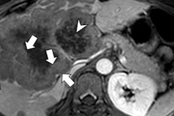
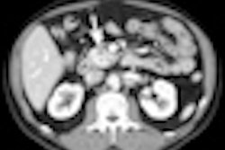
"CT is regarded as a first-line imaging modality for detection and staging of NETs. Thin collimation allows for depiction of millimeter lesions, and multiplanar reconstructions may also help improve lesion assessment," she stated. "The use of biphasic or triphasic contrast-enhanced CT is generally considered a prerequisite for detection and characterization of NETs, because the majority of NET lesions show avid early enhancement. An unenhanced scan can initially be performed to look for calcifications, which for example, occur in around 20% of pancreatic NETs cases, but in only approximately 2% of pancreatic adenocarcinomas."
The basis of functional imaging lies with the targeted detection of specific cell targets or receptors, allowing precise localization of lesions, the authors explained. Somatostatin receptor expression has been found in a large number of tumors, of which NETs are the archetypal class. Somatostatin receptors are widely distributed in the human nervous system and tissues in the body, including the adrenals, kidneys, pancreas, and prostate.
Five subtypes of somatostatin receptors have been identified in humans: SSRT1, SSRT2 -- subtypes 2A and 2B -- SSRT3, SSRT4, SSRT5. Newer generation somatostatin analogs have been developed, allowing radiolabeling with positron-emitting tracers. Together with the development of hybrid PET/CT modalities, several limitations faced with first-generation somatostatin receptor scintigraphy (SRS), largely related to the poorer spatial resolution of gamma cameras and the issue of precise anatomical localization, have been addressed, they noted.
When octreotide is conjugated with the chelator tetraazacyclododecane tetraacetic acid (DOTA), it may be labeled with various radionuclides for imaging and therapy. DOTA-octreotide may be modified with the addition of tyrosine to form DOTA-tyrosine-3-octreotide (DOTA-TOC), which demonstrates a higher affinity for somatostatin receptor subtype 2; or sodium iodide may be added to form DOTA-1-NaI-octreotide (DOTA-NOC), which demonstrates a higher affinity for subtypes 2, 3, and 5, Magalhães pointed out.
"Gallium-68 is a generator-produced positron-emitting nuclide that may be used to label DOTA and its analogs, which allows for high-contrast imaging and detection of small liver, lymph node, and bone metastases and unknown primary tumors in patients who present with metastatic disease," she stated. "With respect to the pancreatic NETs, it is prudent to separate two groups in approaching functional imaging: insulinomas and noninsulinoma pancreatic NETs."
With insulinomas, the role of SRS is uncertain because there is generally poor sensitivity in the detection of such tumors. A significant percentage of insulinomas do not express significant densities of somatostatin receptors, especially subtypes 2 and 5. Moreover, somatostatin receptors are not significantly expressed in nonmalignant insulinomas -- typically only 10% are malignant -- but malignant insulinomas are known to overexpress somatostatin receptors, and SRS has a potential imaging role in such tumors for prognostication and staging. For noninsulinomapancreatic NETs, PET-based SRS has promising utility, according to Magalhães.
NETs arise from cells belonging to the amine precursor uptake and decarboxylation cell system, and therefore show a high 3,4-dihydroxy-L-phenylalanine (L-DOPA) decarboxylase activity, are avid of F-18 DOPA and can be visualized on F-18 DOPA-PET scans. The advantages of DOPA-PET over conventional anatomic imaging or gamma-based SRS are fairly conclusive, but the role in comparison with PET-based SRS techniques is still uncertain, and PET-based SRS appears to be comparable or better in GEP NETs. Moreover, PET-based SRS allows the suitability assessment for peptide receptor radionuclide therapy, something that DOPA-PET does not allow, she concluded.
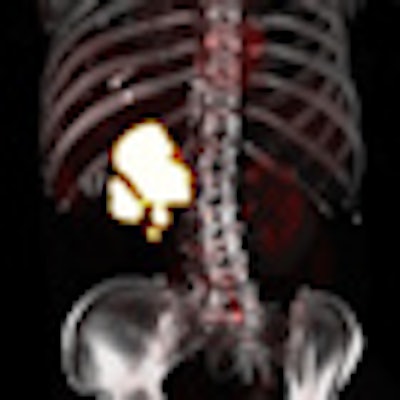
Editor's note: The 3D reconstruction of an abdominal PET/CT scan used on the home page was provided by Dr. Osman Ratib, University Hospital of Geneva, Switzerland.


