A new study from Germany has shown that the eustachian tube opening during a Valsalva maneuver can now be visualized with MRI, suggesting the modality's role in the head and neck region is growing fast. Lesions hampering tubal opening can be delineated during the same MRI examination, and functional MRI of the eustachian tubes may determine the cause of dysfunction.
Real-time imaging has mainly been developed for monitoring of MR-guided interventions, but it can be beneficial in many clinical scenarios, according to Dr. Gabriele Krombach, a professor in the department of diagnostic and interventional radiology at Giessen University Hospital in Giessen, Germany. Depiction of the function of the eustachian tube is just one example.
"We plan to assess real-time imaging for diagnosis of deep vein thrombosis extending from the upper leg into the pelvis in pregnant women. Furthermore, movement of joints can be delineated in real-time, showing moving deficits or hampered movement in patients with injuries," she told AuntMinnieEurope.com. "Another potential indication is assessing cardiac function in arrhythmic patients, in whom ECG triggering is not possible."
Diagnosis of pelvic venous thrombosis is challenging in pregnant women because x-ray exposure should be avoided and ultrasound may be difficult to perform due to the position of the vessels behind the uterus, she explained. With conventional MRI without the use of contrast, it might be difficult to obtain accurate information due to the movement of the unborn child and resulting artifacts.
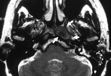 The T2-weighted image shows carcinoma in the left recess of Rosenmüller (arrows). The eustachian tube is not infiltrated, but slightly displaced laterally. All images courtesy of Dr. Gabriele Krombach.
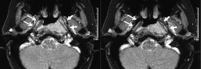 Real-time imaging showed regular opening of the right eustachian tube during Valsalva maneuver (open arrows, right image: neutral position; left image: Valsalva maneuver). The left tube opens only in the anterior part but not in the middle part, due to compression from the tumor (arrows).
Krombach was based at University Hospital, University of Technology (RWTH) in Aachen, Germany when she conducted the head and neck study, along with radiologists Anna Luekens and Dr. Rolf Guenther from RWTH and Dr. Ercole DiMartino from the department of otolaryngology and head and neck surgery at DIAKO Ev. Diakonie-Krankenhaus in Bremen, Germany. The group's work was published online first by European Radiology on 13 October.
The eustachian tube, a connection between the middle ear and nasopharynx, is of great importance in normal ventilation for efficient drainage of the middle ear and hearing. Dysfunction causes symptoms such as hearing disturbances and the feeling of fullness of the ear, and these clinical symptoms can indicate serious diseases such as chronic inflammation of the middle ear or the mastoid, cholesteatoma, or malignant processes affecting nasopharyngeal, inner, or middle ear regions, the authors wrote.
"Owing to its high soft-tissue contrast resolution, MRI represents an excellent method to assess anatomical landmarks and abnormalities in and around the auditory tube. Also, information of eustachian tubes opening and closing, its functional state, is clearly visible with the aid of appropriate protocols while the patients perform a Valsalva maneuver," they stated. "To date, functional studies of the eustachian tube have only been performed in normal subjects, but not in patients with clinical symptoms of nonopening of the eustachian tube and abnormal tympanometry."
Lesions such as nasopharyngeal carcinoma or hyperplastic mucosal swelling, for example, can lead to dysfunction of the eustachian tube so that its physiological tasks are restricted. Without its correct aperture, there is no efficient ventilation, no drainage of the middle ears' secretions, and no protection from excessive sound pressure and pressure differences, all of which can lead to reduced hearing ability, infections, or other middle ear diseases.
Using a 1.5-tesla ACS-NT system from Philips Healthcare, the researchers performed MRI exams on16 patients (seven female, nine male) in the supine position using a standard head coil that was 30 cm in diameter. The age of the patients ranged from 26 to 82 years (mean 55.6 ± 13.1 years standard deviation). Five patients had carcinoma of the neck region, and the remaining 11 patients had clinical signs of sinusitis.
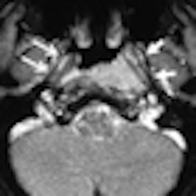
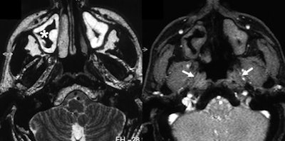 T2-weighting shows sinusitis with swelling of the mucosa. Real-time imaging demonstrates that both eustachian tubes do not open during Valsalva maneuver (right image, arrows).
It was possible to evaluate the opening of the eustachian tube during the Valsalva maneuver in all patients. Within the same examination, an underlying problem was identified and its extent delineated in 14 of the 16 patients. In all patients, the anatomical landmarks and structures were clearly depicted and differentiated from pathological abnormalities in the T2-weighted sequence. Involvement of the anatomical structures could be assessed, but the osseous part of auditory tube was depicted with less good image quality than the other parts. In this region, the amount of soft tissue is small and consists only of the mucous membrane, connective tissue, and the periosteum.
"Visualization of the eustachian tube by MRI constitutes a method unifying imaging, function, and evaluation of pathological findings. Dysfunctionality of the nasopharyngeal tube can be assessed and correlated to its cause in one single examination without any invasive investigations," the authors concluded. "Assessing tubal function is of considerable importance in the preoperative evaluation of patients with chronic otitis media or tympanic effusion, since planning of the operation depends on tubal function. Recovery of tube patency is necessary for prevention of future infections."


