PARIS - Angiography is invasive, costly, and too logistically cumbersome to be used as a diagnostic examination to rule out stenosis. That was the view of Dr. Stephan Achenbach, expressed in a heated debate during the European Society of Cardiology's (ESC) annual congress.
"There are all these smiling people in the cath lab, which is nice, but they have to be paid," he said. "A CT exam reduces staff, takes just a few minutes, and is a great diagnostic test equivalent to angiography for ruling out stenosis."
Achenbach, from the department of cardiology at the University of Giessen in Germany, championed the proposition that CT can replace diagnostic invasive coronary angiography. He was opposed by Dr. Bernhard Meier, director of cardiology at the University Hospital in Bern, Switzerland, who argued that due to its dismal sensitivity, CT is not a useful instrument.
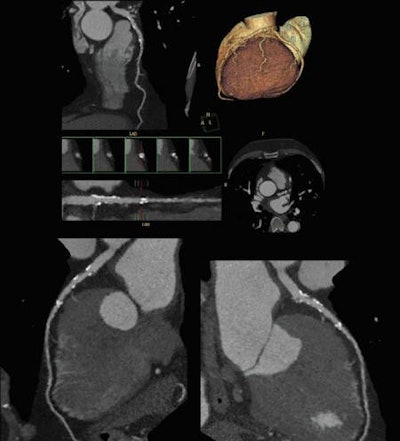
 Cardiac CT angiography performed at lower x-ray dose can help to characterize cardiovascular disease. Image courtesy of Philips Healthcare.
Looking at an occlusion with angiography, you have no idea about what is going on behind that occlusion, but CT can visualize the amount of plaque and also the coronary lumen, according to Achenbach. With a high negative predictive value consistently ranging between 98% and 99% in different studies, CT allows cardiologists to confidently rule out stenosis, giving it a high prognostic value. The modality has a close-to-zero event rate after ruling out stenosis, allowing 84% of patients to be sent home without a single recorded cardiac event among this population over the next 30 days.
"Even though CT has tremendous advantages, I will be the first to admit there are limitations we need to take into account when applying CT to cardiac diagnostics," he said.
CT has lower spatial resolution than invasive angiography, such that severe calcifications can become a problem. Temporal limitations can make motion problematic, requiring a breath-hold and even still producing artifacts within the image due to natural heart movement.
The angiography image may seem misty and blurred, but it shows the whole picture, with a synthesis of all layers of the heart immediately available, Meier commented. It may be old and not fancy, but angiography provides information CT cannot give, such as hemodynamics and flow patterns, and pressure grades can be measured with catheterization, he said.
"The dismal specificity of CT means you are going to have concerns where there is nothing wrong," Meier said. "CT images actually tend to overestimate problems, sometimes dramatically, and you need to look inside the artery to know if there is a clinical significance."
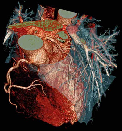
 Cardiac CT images look stunning, but the technique is not perfect. This CT scan used the Flash Spiral Cardio facility of the Somatom Definition system. A 0.9 mSv dose was needed to show the fine details. Image courtesy of Siemens Healthcare.
Meier concedes CT radiation levels have been greatly reduced, but the images produced at 5 mSv do not lead to diagnostic confidence. As a result, patients often undergo a nuclear perfusion study at 9 mSv or possibly even a second examination to validate findings.
"Now we are getting into dangerous radiation levels, and nothing has been done yet," he said. "Why don't we just drop a bomb on this patient?"
All that was needed in the first place was cardiac catheterization, he concluded. "Best of all, invasive angiography is a one-stop shop that includes therapy, so that if we see something of concern, we repair it immediately."


