You could say it's the oldest open "cold case" to date -- the death of the famous Alpine glacier iceman. Since he was discovered in 1991 near the Italy-Austria border, his 5,300-year-old mummified remains have been subjected to numerous scientific tests, and theories have abounded as to how he died. Recently, however, a team of Italian and Swiss researchers determined the iceman's exact cause of death using multidetector-row CT (MDCT).
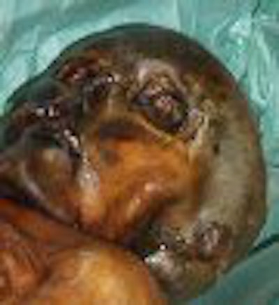
The iceman, known as Ötzi for the Ötztal Valley region where he was discovered, is considered to be the oldest and best-preserved human mummy. Found half-buried in ice by a German couple hiking in the Alps, the corpse was at first believed to be that of an unfortunate modern-day mountain climber. Local officials and a rescue team proceeded to dig him out of the ice (no doubt horrifying crime scene investigators had they been there), and only after examination by an archaeologist was Ötzi's true prehistoric age revealed.
From 1991 to 1998, his remains were housed and studied at the University of Innsbruck in Austria. But after a custody battle of sorts, it was determined that Ötzi was discovered in Italy, and ownership was given to the government of South Tyrol, Italy. Ötzi currently resides in a temperature- and humidity-controlled refrigerated chamber on exhibit at the South Tyrol Museum of Archaeology in Bolzano, Italy.
The study and imaging of Ötzi began with an international team of scientists and researchers from the University of Innsbruck, University of Vienna in Austria, Washington University in St. Louis, University of Texas M. D. Anderson Cancer Center in Houston, and General Regional Hospital in Bolzano, Italy.
Scientific wonder
Since his discovery, Ötzi has fascinated the scientific community, as his body was found frozen and well-preserved. The clothing he wore at the time of his death also survived, as well as his tools and other accoutrements, giving scientists a glimpse into life during the Neolithic-Copper Age.
Clothing items found include his goat-hide coat and leggings, leather loincloth, calf-leather belt with a pouch, bear- and deerskin shoes stuffed with hay, bearskin cap, and remnants of an Alpine grass cloak. He also had with him a backpack frame, copper-blade axe, quiver and arrows, unfinished bow, flint, dagger, grass net, and even a "medicine kit" of birch polyporus, perhaps for its antibiotic and styptic properties.
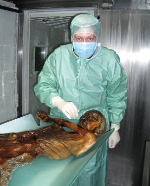 |
| Dr. Frank Rühli examines the Iceman. Image courtesy of Dr. Frank Rühli and the University of Zurich, Switzerland. |
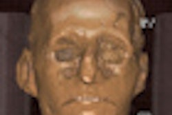
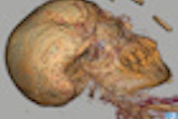
From head to toe, Ötzi has been subjected to numerous tests, including Raman spectroscopy and chromatography of his skin, electron microscopy of lung particles, bone scans, and DNA testing, by scientists across the globe. His skull has been studied and reproduced with CT-guided stereolithography. Using 3D computer-assisted biopsy, samples were taken from his nose, maxillary sinus, and larynx. Other samples of his ribs, blood vessels, liver, spleen, diaphragm, femoral muscle and nerve, and brain were taken endoscopically. The contents of his stomach and colon were examined -- his last meal consisted of red deer meat and grains. Even the construction and materials of his footwear have been studied, inspiring modern reproductions.
After all this examination, researchers believe Ötzi died at the approximate age of 45. He weighed about 99 lb and stood about 5 feet, 2 inches tall (Experimental Gerontology, November-December 1998, Vol. 33:7-8, pp. 655-60).
Imaging the iceman
As imaging technology has evolved, so has the imaging scans that the iceman has undergone. In 1991, the mummy was scanned with conventional CT (Somatom Plus, Siemens Medical Solutions, Erlangen, Germany) with sections of 1-8 mm. In 1993, the remains underwent CT scanning again, but this time with a portable unit to limit time in transit and to minimize thawing. A spiral CT scanner (Somatom Plus 40, Siemens) was used in 1994 in Austria and again in 2001 in Italy, and digital radiography was also used in 2001.
"In all, 38 radiographic and approximately 2,190 CT source images were obtained for interpretation. Many two- and three-dimensional reconstructed images were derived from the CT datasets. These were used to gain additional anatomic perspective and to aid anthropologists and other investigators in their studies," wrote Dr. William Murphy Jr., from the division of diagnostic imaging at the M. D. Anderson Cancer Center in Houston, and colleagues (Radiology, March 2003, Vol. 226:3, pp. 614-629).
Besides the injuries that occurred from being frozen beneath a glacier for ages and dug out of the ice with tools such as ice axes, ski poles, sticks, rocks, and a pneumatic jackhammer, Ötzi was shown to have healed rib fractures of the fifth through ninth ribs on his left side, degenerative arthritis in his spine and left hip, and vascular calcifications in the carotid arteries, distal aorta, and right iliac artery -- not to mention a case of frostbite on his left little toe.
However, "bone mineral content was excellent, and the lower-extremity long-bone cortices were particularly thick," Murphy's group wrote. "Thick cortices were associated with prominent nutrient canals and attachments for muscles (linea asperae) and tendons (tibial tubercles)." A life spent climbing and hiking in the Alps favored the development of muscle and bone, they explained.
A common theory as to how Ötzi died was that he got caught in a storm and froze to death. But the 2001 digital x-ray findings suggested otherwise. X-rays of the ribs showed an arrowhead lodged between the rib cage and left scapula, indicating that he had been shot in the back and leading researchers to re-examine previous CT images.
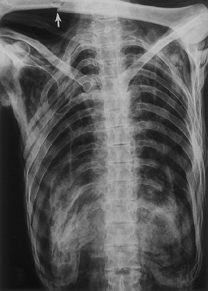 |
| Frontal radiograph of the chest and upper abdomen (obtained on May 25, 1993) confirms the caudal rotation of the ribs and shows the folded soft tissues at the thoracolumbar junction. Only 11 rib-bearing thoracic vertebrae are present. The usual shadows of the heart, pulmonary vessels, and lungs are absent because of severe shrinkage of these organs. The fracture (arrow) of the left humerus was acquired during the recovery effort. |
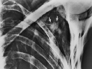 |
| Irregular opacity (arrows) in the left shoulder retrospectively detected on a conventional frontal radiograph of the chest (obtained on September 26, 1991) has the general configuration of an arrowhead. Also in retrospect, the arrowhead is visible on the radiograph obtained on May 25, 1993 (top). Fig. 8, 21, Murphy WA, zur Nedden D, Gostner P, et al. "The Iceman: Discovery and Imaging," Radiology 2003; 226:614-629. |
The CT images also displayed hyperattenuating areas that were interpreted as dehydrated remnants of a hematoma, as well as evidence of a perforation of the scapula, the researchers stated. After inspecting the mummy's back when the arrowhead was discovered, the researchers found a small skin laceration over the scapula. They hypothesized that the arrow entered the left shoulder from the rear, passed through the scapula, and injured a major vessel, resulting in a hematoma.
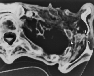 |
| Transverse CT section (obtained on May 3, 1994) through the left shoulder region (caudal to the prehistoric arrowhead) shows an inhomogeneous area of attenuation (dehydrated hematoma, arrow) between the lateral rib cage and the scapula. Note the discontinuity in the ossified body of the scapula and the wispy soft-tissue opacity (hematoma) that extends through the bone defect (arrowheads). Authors speculate that the arrowhead and a portion of the arrow shaft penetrated the scapula in this location and that blood from the deep hematoma followed the arrow track into the subcutaneous tissues. Fig. 23, Murphy WA, zur Nedden D, Gostner P, et al. "The Iceman: Discovery and Imaging," Radiology 2003; 226:614-629. |
"Furthermore, it is speculated that when the arrow was withdrawn, the overlying scapula interfered and caused the arrowhead to separate from the shaft. Thus, the arrowhead remained trapped between the rib cage and the scapula," Murphy and colleagues wrote. "The fact that the arrow entered the iceman from behind suggests that the manner of death was either accidental or homicide."
MDCT: Case closed
More recently, researchers used multidetector-row CT (MDCT) to radiologically prove Ötzi's exact cause of death, expanding on the previous findings. Their results were published online in the Journal of Archaeological Science and in the July issue of National Geographic.
In August 2005, the mummy was re-examined in South Tyrol, Italy, by Dr. Frank Rühli of the Institute of Anatomy at the University of Zurich in Switzerland and colleagues from the departments of radiology and pathological anatomy and histology at General Hospital Bolzano in Balzano, Italy.
Rühli is a co-chair of the Swiss Mummy Project. The project's goal is to use noninvasive methods, such as CT and other radiological techniques, to obtain information on historic mummies. The project is funded by the University of Zurich, as well as Siemens, the Zuse Institute Berlin in Germany, and the Reiss-Engelhorn Museum in Mannheim, Germany. Rühli and some of his colleagues were also consultants in determining the cause of death of Egyptian pharaoh Tutankhamun in 2005.
The group examined the iceman's perimortem subclavicular vascular lesions using a 16-detector-row CT scanner (Sensation 16, Siemens) with slice collimation of 0.75 mm, table increment of 6 mm, 120 kV, and 180 mAs. Multiplanar reformatting and volumetric 3D imaging were performed on a Leonardo workstation (Leonardo Leo Syngo2004A VD10B, Siemens).
Using MDCT, the researchers were able to identify the subclavian artery with its higher density (-40 HU), compared to the surrounding soft tissue (-290 HU), and found damage to the left dorsal artery wall that was 13 mm in length. Also visible was a 3-mm irregular pseudoaneurysm, which they noted was a typical complication of a subclavian artery laceration. A large hematoma was seen in the surrounding soft tissue, and the arrowhead (+1840 HU) was found in situ 6.5 mm away in the dorsocranial direction, they added.
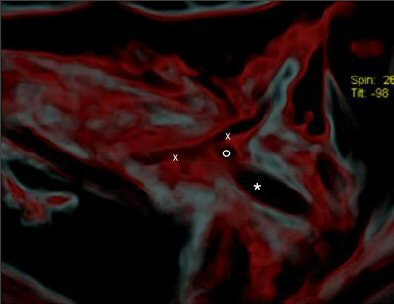 |
| CT image shows the injury (x-x) and the head of the arrow (*). Figure published in: JArcheol Sci, in press, Pernter P., Gostner P., Egarter Vigl E., Rühli, F. J., "Radiologic proof for the Iceman's cause of death (ca 5300 BP), Copyright Elsevier. Image courtesy of Dr. Frank Rühli and the University of Zurich, Switzerland. |
"It seems most likely that (the arrowhead) lacerated the subclavian artery. By removing the shaft of the arrow perimortem, its head must have been slightly retracted to the actual position where the barbs caught in the tissue and, eventually, the arrowhead separated from the now missing shaft," the group wrote. "In the surrounding soft tissue one can see linear air incorporations as well as multiple irregular partially confluent densities (-80 HU), with the latter likely representing a hematoma. It spreads dorsocaudally between the ribs and the scapula, and also into the shooting channel toward the subcutaneous soft tissues" (Journal of Archaeological Science, March 15, 2007).
The researchers noted that major symptoms of subclavian artery injuries often include massive active bleeding, expanding hematoma, and shock-related cardiac arrest. "The apparent lack of intravascular blood and the lack of postmortem lividity (livor mortis) in the iceman fit well with the diagnosis of injury-related complete perimortem exsanguination," they wrote.
"The iceman's cause of death by an arrowhead lacerating, among others, a great thoracic artery -- the left subclavian artery -- and leading to a deadly hemorrhagic shock can be now postulated with almost complete certainty, especially when considering environmental (3,210 meters above sea level) and historic (5,300 BP [before present]) settings into account," the researchers concluded.
By Heather Hokenson
AuntMinnie.com staff writer
July 4, 2007
Related Reading
Mystery of King Tut's demise solved? November 29, 2006
Imaging reveals truth about Barnum mummy, October 31, 2006
Scan artist: Radiologist uses CT to reveal mystery of antiquities, October 25, 2005
CT helps unwrap mummy mystery, March 29, 2005
MDCT solves 400-year-old mystery, November 29, 2004
Copyright © 2007 AuntMinnie.com


